


When Aching Below the Kneecap Comes Back in Adulthood — Do Not Confuse ‘Adult Osgood-Schlatter Sequelae’ with Patellar Tendinopathy or Knee Osteoarthritis: Dr. Moriwaki Explains the Chronic Inflammation Cycle Around the Retained Ossicle at the Tibial Tuberosity, and Where Peri-Knee Injection of Stem Cell Conditioned Media Can Calm the Inflammatory Environment — and Where It Cannot Physically Remove the Bone Fragment Itself2026.08.14
“I remember being diagnosed with Osgood-Schlatter disease as a teenager, but back then the pain settled within several months to a year” — patients who remember it this way sometimes find, in their 30s or 40s, that the area just below the kneecap (the tibial tuberosity) has started to ache again. When the orthopedist diagnoses “patellar tendinopathy” or “knee osteoarthritis” and the symptoms still refuse to resolve, adult Osgood-Schlatter sequelae may be hiding in the background. Adult Osgood-Schlatter sequelae is a condition in which the tibial tuberosity apophysitis of adolescence did not fully heal, an ossicle (bone fragment) remained after the growth plate closed, and in adulthood that fragment and the patellar tendon insertion continue to grind against each other and generate a chronic cycle of inflammation. In this article, Dr. Moriwaki lays out the pathology, the differential diagnosis points, and where peri-knee injection of stem cell conditioned media can and cannot help — the boundary between calming the inflammatory environment and physically removing the bone fragment itself.
Key Points of This Article
・Adult Osgood-Schlatter sequelae is a condition in which the ossicle left behind by adolescent tibial tuberosity apophysitis continues to rub against the patellar tendon insertion in adulthood, producing a chronic inflammatory cycle.
・Because the pain lies close to that of patellar tendinopathy (jumper’s knee) and knee osteoarthritis, misdiagnosis is common; the presence of localized tenderness over the tibial tuberosity is the gateway to distinguishing them.
・Diagnosis rests on history (past Osgood in adolescence), palpation of the tibial tuberosity, and imaging (ultrasound, X-ray, MRI) to visualize the ossicle and tendon insertion.
・Peri-knee injection of stem cell conditioned media may be considered to calm the inflammatory environment of the enthesis, but it cannot physically remove the bone fragment itself.
・When the fragment is large and mechanical irritation dominates, surgical ossicle excision may take priority over conservative care.
What Is Adult Osgood-Schlatter Sequelae as a Pathology?
Why the ossification does not fully unite in adolescent Osgood
Osgood-Schlatter disease occurs around ages 10–15, when the secondary ossification center at the tibial tuberosity (the bony bump just below the kneecap) is stressed by the pull of the patellar tendon during a growth spurt, causing partial avulsion and incomplete ossification. In most people, symptoms resolve naturally as the growth plate closes at around 16–18, but sometimes the small avulsed fragment does not fuse with the main tibia and remains as an independent free ossicle. This fragment sits within or at the patellar tendon insertion (enthesis), and every time the tendon stretches and contracts — with squatting, stair climbing, jumping — the surrounding tendon tissue and bursa take on minute mechanical stress. Even if there were almost no symptoms in youth, aging brings reduced tendon elasticity, and mid-life shifts in activity can trigger pain to surface as adult Osgood-Schlatter sequelae.
A palpable bony bump at the tibial tuberosity — and localized tenderness
Many patients with adult Osgood-Schlatter sequelae can feel a distinct bony bump at the front-lower aspect of the knee (the tibial tuberosity, just below the kneecap). This is the ossified remnant of the fragment retained during adolescence. Pressing on this bump reproduces a sharp, well-localized tenderness, and deep knee flexion spreads an aching sensation around the area. It is also common for symptoms to flare after resuming sports that involve rapid knee flexion-extension (long-distance running, kendo lunges, futsal, tennis) or after prolonged squatting work.
Differentiating Adult Osgood Sequelae from Patellar Tendinopathy and Knee Osteoarthritis
Difference from jumper’s knee (patellar tendinopathy)
So-called jumper’s knee (patellar tendinopathy) is characterized by pain centered at the “lower pole of the patella.” In contrast, the tenderness of adult Osgood-Schlatter sequelae concentrates lower, at the tibial tuberosity. Jumper’s knee involves chronic degeneration at the patellar origin of the tendon; Osgood sequelae involves inflammation at the tibial insertion of the tendon plus the surrounding retained ossicle — the “address” of the source is different. Scanning the patellar tendon from the lower pole of the patella down to the tibial tuberosity with ultrasound clearly separates the two, and matching the pain location to imaging carefully is the key to raising diagnostic accuracy.
Difference from knee osteoarthritis
The pain of knee osteoarthritis (knee OA) typically spreads across the medial joint line, the patellofemoral joint, and the joint as a whole, worsening with descending stairs and rising from a chair. Adult Osgood sequelae keeps the pain localized to the tibial tuberosity, below the joint line, and worsens with deep flexion (kneeling, deep squatting). Since age often brings knee OA as a comorbidity, when both coexist the pattern “knee OA medication keeps the joint quiet but the pain just below the kneecap remains” is a common way this sequela gets noticed. Distinguishing the individual pain sources requires combining physical examination with imaging.
Where Peri-Knee Injection of Stem Cell Conditioned Media Can Reach — and Where It Cannot
What it can target: calming the inflammatory cycle at the tendon insertion
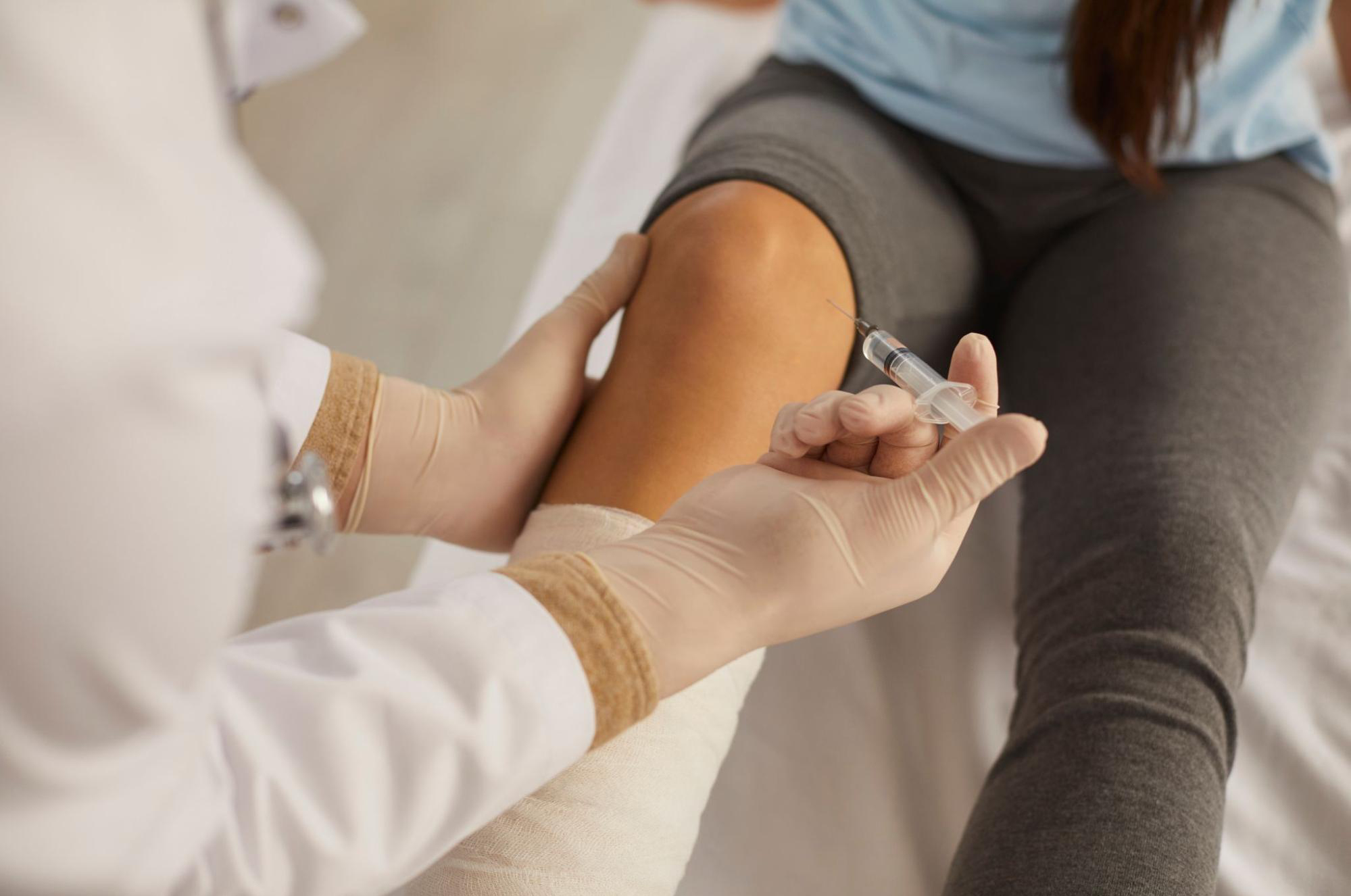
The chronic pain of adult Osgood sequelae is driven by a small-scale inflammatory cycle repeating between the retained ossicle at the tibial tuberosity and the patellar tendon insertion. The tendon insertion (enthesis) is known as a site with poor blood supply and slow tissue repair, and ordinary oral NSAIDs and icing often struggle to settle it. Stem cell conditioned media contains growth factors such as TGF-β, IGF-1, and FGF, along with anti-inflammatory cytokines, and can be expected to act on the inflammatory environment of the tendon insertion. Delivering it under ultrasound guidance as a peri-knee injection close to the tendon insertion at the tibial tuberosity aims to lower the pain baseline so that rehabilitation can be continued more consistently. That said, response varies by individual, and not every case will show the same reaction.
What it cannot target: physically removing the bone fragment itself
On the other hand, injecting stem cell conditioned media is a pharmacological action; it cannot dissolve or physically remove the bone fragment that remains at the tibial tuberosity. When the fragment is large (for instance, 10 mm or more in diameter) and clearly obstructs the course of the patellar tendon, or when mechanical irritation keeps flaring symptoms despite months of conservative care, surgical ossicle excision becomes the priority option. Making this boundary clear from the first visit is essential so that the patient can realistically judge “how much the injection can be expected to do.”
How Other Conservative Treatments Are Combined
Conservative care for adult Osgood-Schlatter sequelae is designed in stages: (1) activity modification and stretching of the quadriceps and hamstrings; (2) physical therapy (ultrasound, extracorporeal shockwave); (3) analgesics (oral or topical NSAIDs); (4) bracing (patellar tendon strap); (5) local injections (steroids, hyaluronic acid, stem cell conditioned media); and (6) surgical ossicle excision for severe cases with a poor response. Local steroid injection can bring short-term relief, but repeated injections into the tendon itself carry a known risk of tendon weakening and require careful dosing. Stem cell conditioned media differs from steroid in that its purpose extends beyond suppressing inflammation to correcting the tissue repair environment; it is one conservative option chosen with a view to calming the chronic inflammatory cycle of the tendon insertion over the medium to long term. For general information on joint disease, you may also refer to the website of the Japanese Orthopaedic Association.
For details on our treatment design and indications, please also see more information on joint injection with stem cell conditioned media.
Frequently Asked Questions
Q. Can Osgood pain really flare up again in adulthood?
Yes — it is a condition experienced by a certain proportion of patients as adult Osgood-Schlatter sequelae. The retained fragment at the tibial tuberosity from adolescence, together with age-related reduction in tendon elasticity or a mid-life return to sports, can trigger a chronic inflammatory reaction through friction with the surrounding patellar tendon insertion. The first step is to check whether the recalled history and the current location of tenderness at the tibial tuberosity match.
Q. How many peri-knee injections of stem cell conditioned media are typically needed?
The plan varies with individual symptoms and response, but a common starting point is several injections spaced 4–8 weeks apart during the induction phase, followed by an assessment of effect and adjustment of intervals for maintenance. Because chronic inflammation at the tendon insertion takes time to improve, combining multiple sessions with parallel stretching and activity control is important, rather than expecting a single injection to be enough.
Q. If a fragment is left behind, will surgery to remove it lead to complete cure?
When the fragment is large and clearly the primary source of mechanical irritation, ossicle excision can significantly improve symptoms. However, surgery requires post-operative tendon healing and rehabilitation, and it is not the first-line choice for every case. The usual stepwise approach is to try conservative care (stretching, bracing, local injection) for several months first, and consider surgery when the response is poor.
Q. Can I go back to running and sports right after the injection?
For about 72 hours after the injection, avoid intense exercise; once swelling and warmth settle, gradually return to activity starting with light loads. Because the injection is meant to calm chronic inflammation, immediate overload can worsen symptoms. The timing of return to competition should be judged individually by objectively assessing pain, range of motion, and recovery of quadriceps strength.
Q. I also have knee osteoarthritis. Can both be treated at the same time?
Because knee OA and adult Osgood-Schlatter sequelae have different pathologies and therapeutic targets, they are usually treated in parallel. One combination is intra-articular injection for knee OA and peri-tendon-insertion injection for Osgood sequelae — administering stem cell conditioned media at different target layers. Individual joint status should be evaluated with X-ray, MRI, and ultrasound before setting priorities.
──────────────
Supervising Physician: Dr. Shin Moriwaki
Member, Japan Society of Aesthetic Surgery (JSAS) / Member, American Academy of Aesthetic Medicine
ECFMG Certificate
──────────────
📍AVAN TOKYO 銀座 関節再生医療
AVAN TOKYO Ginza Joint Regenerative Medicine
English / 中文 / Tiếng Việt available
Inquiries welcome via DM / LINE / Website / Phone.