


When Three Joint Injections Fail: Rethinking Diagnosis and Next Steps2026.10.20
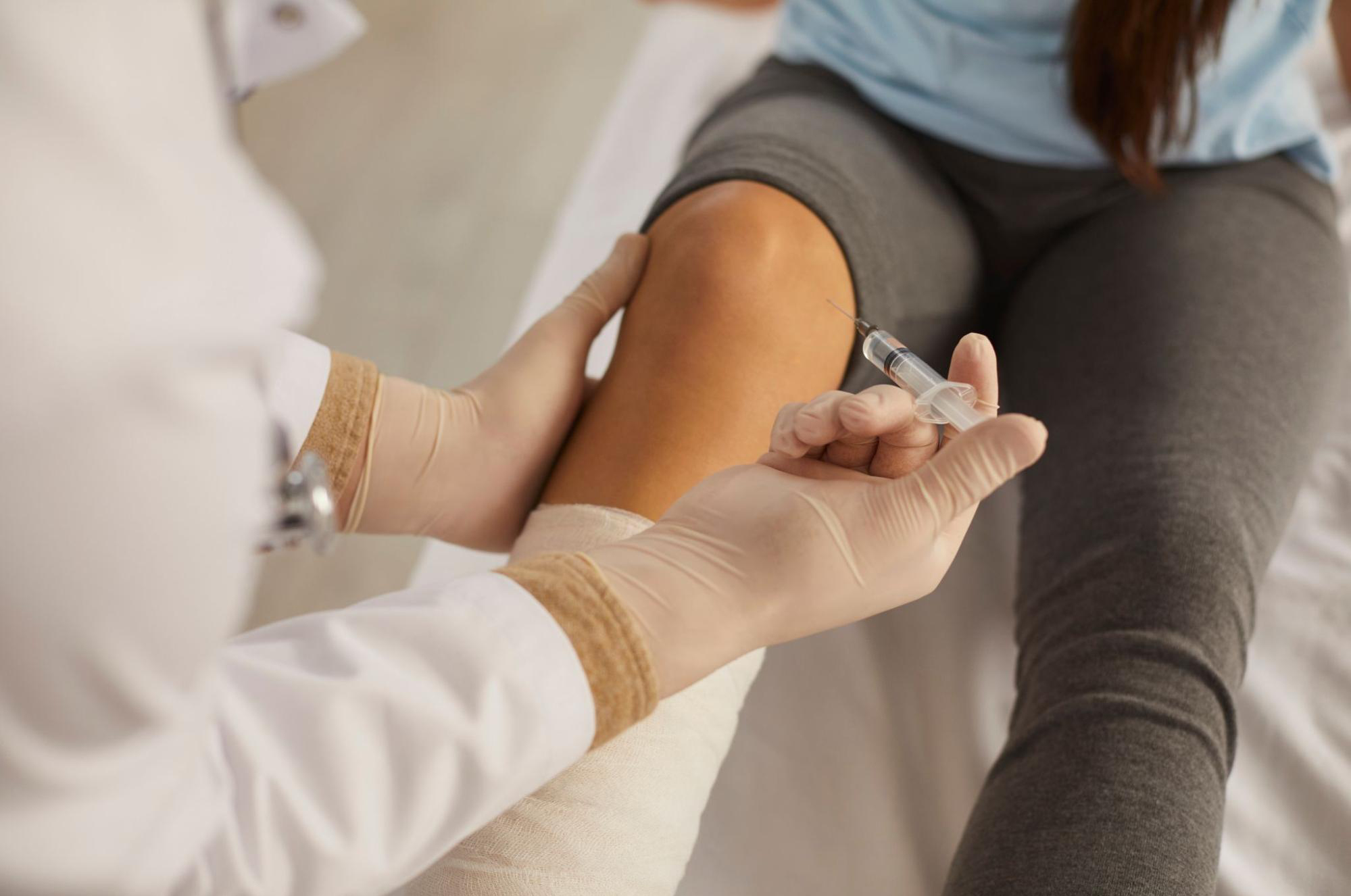
Many patients with knee osteoarthritis or adhesive capsulitis of the shoulder complete three joint injections only to say “the pain is unchanged” or “my range of motion has not come back.” Before concluding joint injection poor response, what truly needs re-examination are two things: the diagnosis itself, and the targeting accuracy of the injection. In this article, Dr. Moriwaki walks through what orthopedic specialists re-evaluate when a three-injection course produces little change, which additional investigations to consider, and the next-line options including stem cell conditioned media injection and exercise therapy redesign.
Key points of this article
・If you suspect joint injection poor response, the first priority is to question whether the original diagnosis is still valid.
・When hyaluronic acid or steroid injections produce no change after three attempts, the pain generator may not match where the needle is being placed.
・Synovitis, bone marrow edema, and enthesopathy are often invisible on plain X-ray and should be re-evaluated with MRI or ultrasound.
・The four axes for your next step are: changing the drug, switching to ultrasound-guided puncture, trying stem cell conditioned media, and redesigning exercise therapy.
・Do not self-decide that “three times means it’s over.” Keep a record of the course and update the plan with your physician.
Three injections without effect is not unusual
Joint injection is not a treatment that reaches a conclusion in a single session. Clinicians usually treat two to three injections as one evaluation window and read the response from there. Even so, 30 to 40 percent of patients in real-world practice report “no change” or “relief only the next day, then back to baseline.” The reasons for non-response fall into three major categories.
1. The diagnosis itself is off
A label of knee osteoarthritis does not guarantee that OA is the actual pain generator. The true source may be a medial meniscus posterior root tear, pes anserine bursitis, patellofemoral arthrosis, or medial plica syndrome. Even with Kellgren-Lawrence grade II or III changes on X-ray, the real culprit may lie elsewhere. If the inflammatory origin is in extra-articular tendons or bursae, intra-articular injection will respond poorly.
2. The injection misses the target layer
Blind (landmark-based) puncture achieves intra-articular placement in only 70 to 80 percent of attempts for some joints. The hip, subscapular bursa, and posterior ankle are particularly hard to confirm by surface landmarks, and when the drug spreads outside the joint, the effect is halved. Targeting accuracy is a major reason the same drug produces different responses in different hands.
3. The drug does not match the pathology
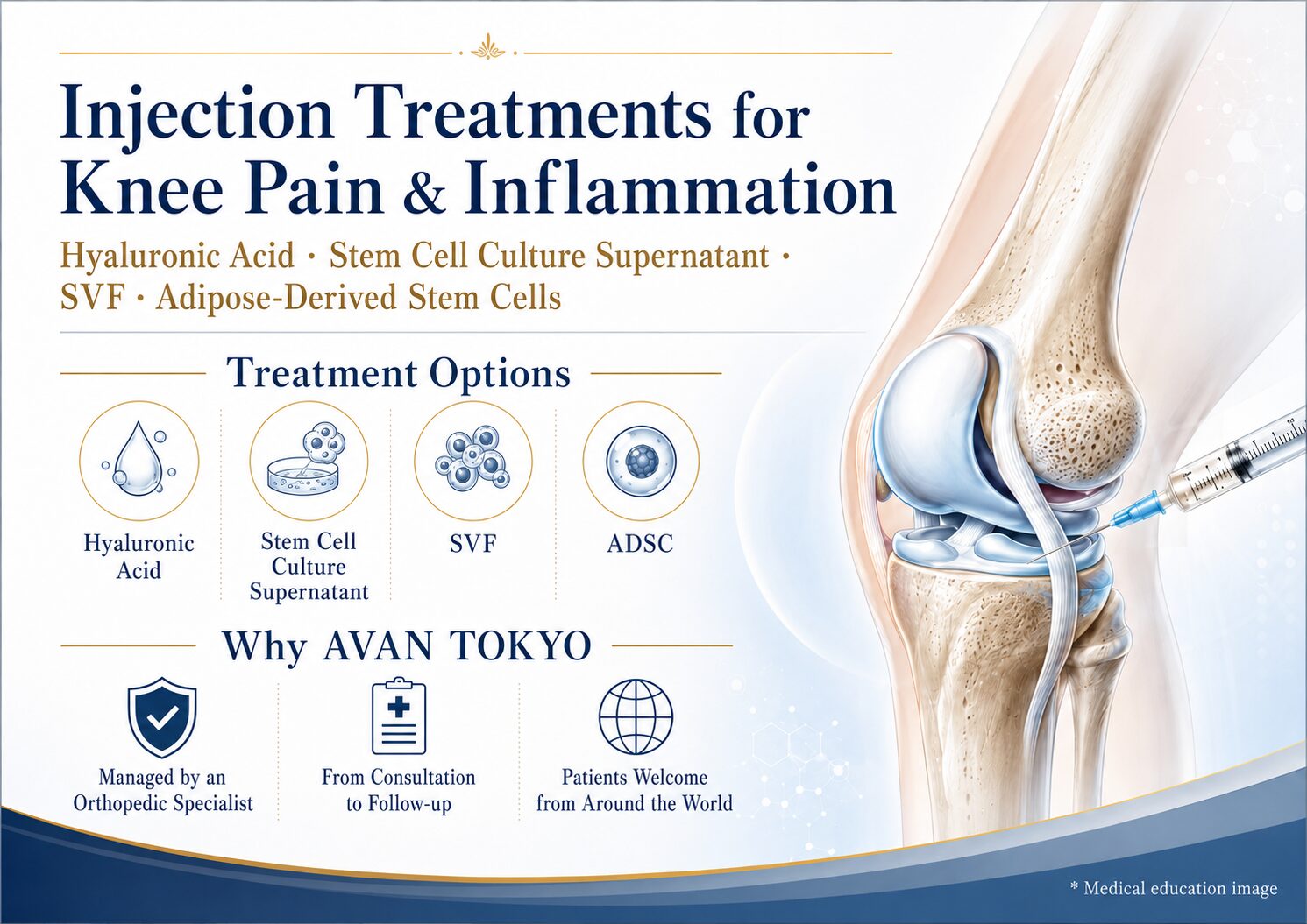
Hyaluronic acid replenishes viscoelasticity, steroids suppress strong inflammation, and stem cell conditioned media modulates the intra-articular cytokine environment. Mechanisms differ. Hyaluronic acid alone for acute synovitis, or repeated steroid for chronic tendinopathy, both tend to produce poor responses. A pathology-drug mismatch is a classic reason for “three injections and no effect.”
Four axes to revisit when you suspect joint injection poor response
Reconfirm the diagnosis
If the initial diagnosis was based on X-ray alone, consider MRI. Meniscus posterior root tears, bone marrow edema, synovitis, and enthesopathy are not seen on plain films. If possible, add ultrasound dynamic assessment, moving the joint while identifying where pain arises, to separate intra-articular from extra-articular pain. In the interview, also recheck whether pain is at rest or on motion, whether night pain is present, and whether the patient can point to one spot with a single finger.
Revisit the puncture technique
Simply switching to ultrasound guidance can improve drug delivery accuracy and change outcomes with the same medication. The knee alone has four puncture approaches, chosen according to the presence of effusion and the position of synovial plicae. The harder the joint is to localize by surface landmarks, as in the hip, posterior ankle, or subscapular bursa, the greater the gain from ultrasound.
Redesign drug selection
If steroid has not worked after three attempts, respect the annual injection limit and switch tracks. If hyaluronic acid has produced no change, consider stem cell conditioned media or extracorporeal shockwave, which address the inflammatory cytokine environment. Stem cell conditioned media is not a cartilage-regenerating treatment per se; the concept is to break the inflammatory loop by modulating the intra-articular environment. Please also see more about stem cell conditioned media joint injection here.
Exercise therapy, weight, and posture
Without quadriceps strength, body weight management, and gait correction, no injection sustains its effect. Any pain that cannot be solved by injection alone is almost always accompanied by extra-articular load factors. A 1 kg increase in body weight translates to several kilograms of load on the knee during walking, so weight, strength, and alignment must be reviewed in parallel with injection therapy.
Choosing the next step
Option A: change the drug
No change with hyaluronic acid → short-term steroid introduction, or combination of hyaluronic acid with stem cell conditioned media. If repeated steroid is losing effect, respect the three to four annual injection cap and consider stem cell conditioned media or shockwave therapy from the next session.
Option B: change the technique
If three blind injections have not produced change, switch to ultrasound-guided puncture. Re-select the puncture site (suprapatellar pouch, inferolateral, or medial border), and when effusion is present, aspirating before injection often changes the response.
Option C: inject outside the joint
If intra-articular injection does not work, redirect to extra-articular pain generators such as pes anserine, medial collateral ligament insertion, patellar tendon, or greater trochanter. For general information on joint disease, the Japanese Orthopaedic Association is also a useful reference.
Option D: re-evaluate surgical indication
If MRI reveals a meniscus posterior root tear, idiopathic osteonecrosis, or end-stage subchondral change, having orthopedic surgery evaluated earlier may reach relief faster than continuing injections. Joint injection is a cornerstone of conservative care, but it is not universal, and recognizing the border with surgical indication is essential.
FAQ
Q. After how many ineffective injections should I give up?
The threshold for joint injection poor response is three consecutive sessions with the same drug at the same site producing little change. The right next step, however, is not “to give up” but “to re-examine diagnosis and technique.” Prioritize changing the axis of evaluation over simply increasing the number of injections.
Q. Do three steroid injections damage cartilage?
Intra-articular steroid injections exceeding three or four times a year are reported to carry risks of cartilage impact and elevated blood glucose. If three consecutive injections produce little effect, switching tracks rather than increasing the dose is the sound judgment.
Q. How many sessions do I need to judge stem cell conditioned media?
As an induction course, three sessions at three to four week intervals is a common baseline for reading response. If the response remains limited, concentration, site, and combination therapy are reviewed. Like hyaluronic acid, it is not a treatment that decides in one session.
Q. Does it make sense to continue the same injection that did not work at another clinic?
In principle, the value is limited. Repeating injections without changing diagnosis or technique only adds intra-articular load while plateauing the benefit. Reviewing imaging, technique, and drug simultaneously is the realistic approach.
Q. When should I seek a second opinion?
If three injections have brought little change and the next plan is simply “keep doing the same injection,” consulting another facility to review diagnosis, imaging, and technique is realistic. Bringing a course memo, imaging data, and the names of medications used will accelerate the discussion.
──────────────
Supervising physician: Shin Moriwaki, MD
Member, Japan Society of Aesthetic Surgery (JSAS) / Member, American Academy of Aesthetic Medicine
ECFMG Certificate
──────────────
📍AVAN TOKYO Ginza Joint Regenerative Medicine
AVAN TOKYO Ginza Joint Regenerative Medicine
English / 中文 / Tiếng Việt available
Inquiries welcome via DM / LINE / Website / Phone.