


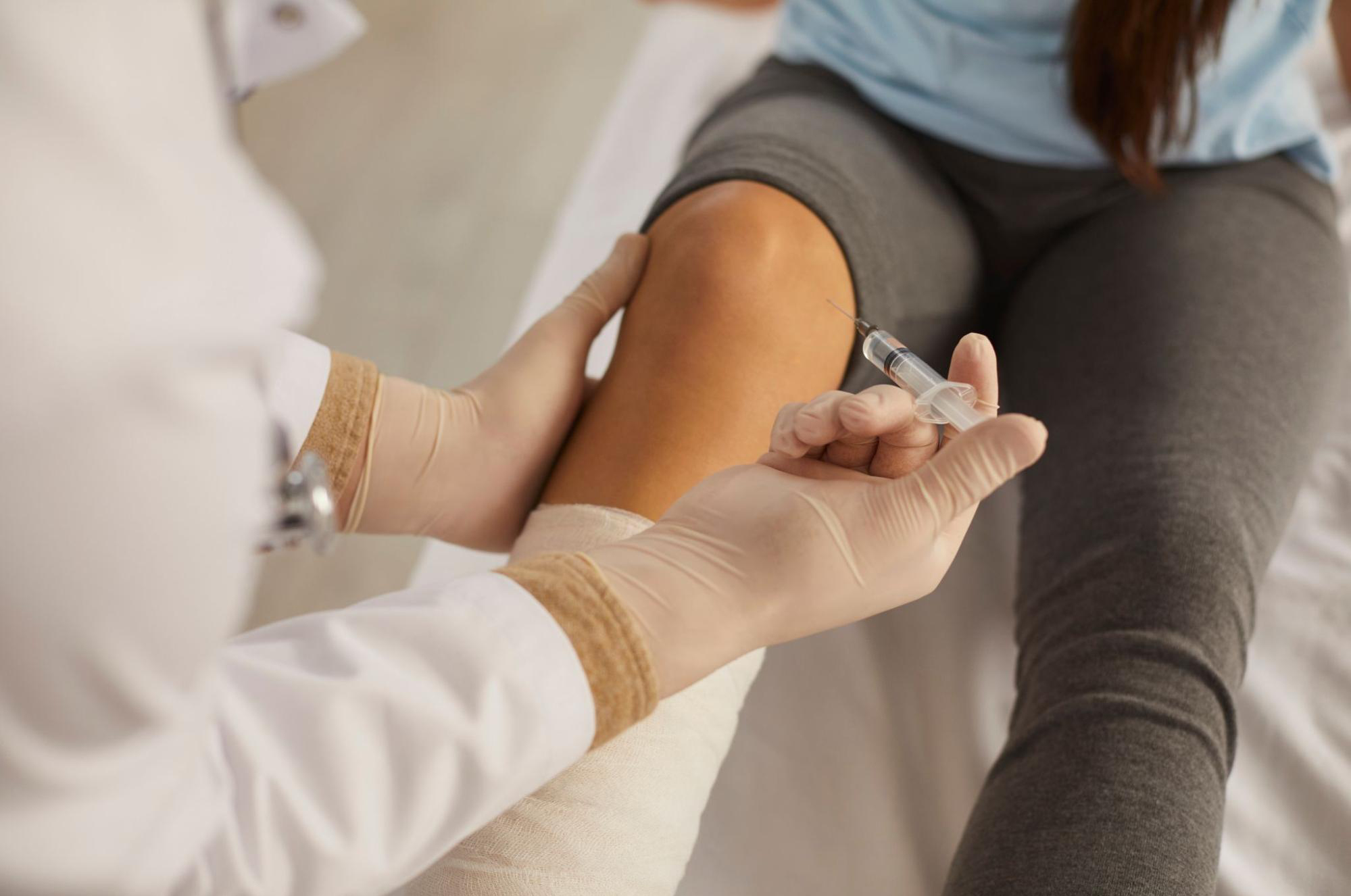
Don’t dismiss ‘medial meniscus extrusion’ on your knee MRI as ‘just aging’: how a meniscus pushed outside the joint line accelerates cartilage loss, and where stem cell conditioned media knee injection can help address intra-articular inflammation versus where it cannot restore anatomical position — organized by Dr. Moriwaki2026.08.16
When a knee MRI report reads ‘medial meniscus extrusion,’ patients are often told ‘it’s an age-related finding.’ But is that really the whole story? Recent osteoarthritis research shows that medial meniscus extrusion is not merely a benign aging change — it is now recognized as an important predictor of how quickly knee osteoarthritis (KOA) will progress. In this article, Dr. Moriwaki honestly explains why medial meniscus extrusion drives a joint-damaging cycle, what stem cell conditioned media knee injection can realistically address, and what structural limits injection cannot cross.
Key Points of This Article
・Medial meniscus extrusion is a state in which the meniscus is pushed outside the joint line, reducing the contact area of cartilage and accelerating knee osteoarthritis
・Observational studies suggest that extrusion beyond 3 mm significantly accelerates medial compartment cartilage wear, so it should not be brushed aside as ‘just aging’
・Most cases coexist with medial meniscus posterior root (root ligament) injury or degenerative meniscus changes, and root tears themselves cannot be sutured by injection
・Stem cell conditioned media knee injection targets the correction of the intra-articular pro-inflammatory cytokine environment, not the anatomical repositioning of an already extruded meniscus
・Diagnosis is centered on MRI coronal images rather than plain X-ray, and treatment design must integrate injection, bracing, exercise therapy, and surgical indication based on alignment, muscle strength, and KL grade
What Is Medial Meniscus Extrusion — What the MRI Actually Shows
Meniscal anatomy and ‘push-out beyond the joint line’
The meniscus is a crescent-shaped fibrocartilage between the femur and the tibia that distributes load and assists joint fluid lubrication. A healthy meniscus sits inside the medial joint line, but as age-related tissue degeneration and chronic mechanical loading accumulate, its ligamentous support weakens and it is gradually pushed outside the joint space. This condition is termed ‘medial meniscus extrusion,’ and it is measured on MRI coronal images as the distance from the tibial articular edge to the outer edge of the meniscus.
Beyond 3 mm signals accelerated progression
Multiple observational studies report that when extrusion exceeds 3 mm, medial compartment cartilage wear accelerates and progression along the KL classification speeds up. That said, extrusion width fluctuates between weight-bearing and non-weight-bearing conditions, so it is unwise to conclude from a single static image; symptoms, examination findings, and multiple imaging modalities must be evaluated together.

Why Medial Meniscus Extrusion Accelerates Knee Osteoarthritis
Reduced contact area and stress concentration
The meniscus distributes body weight over a broad cartilage contact area, lowering stress per unit area. Once the meniscus is pushed out, however, the cushioned zone inside the joint shrinks, and the medial femoral condyle and medial tibial condyle come into near-‘bare contact.’ Higher unit-area stress accelerates cartilage matrix degradation, bone marrow edema, and subchondral micro-fractures — clinically expressed as medial-side pain worsening on stair descent, prolonged walking, or seiza (kneeling).
Continuity with root tears and the synovitis loop
Most cases of medial meniscus extrusion occur in continuity with medial meniscus posterior root tears. When the root ruptures, the meniscus loses its ‘rubber-band’ hoop function, so every weight-bearing cycle actively pushes it outward. Debris and mechanical abrasion from the torn meniscus sustain chronic synovitis inside the joint, and pro-inflammatory cytokines such as IL-1β and TNF-α tilt cartilage metabolism toward degradation, accelerating extrusion in a vicious loop.
What Stem Cell Conditioned Media Knee Injection Can and Cannot Target
What it can address — correcting the intra-articular inflammatory environment
Stem cell conditioned media contains anti-inflammatory cytokines and multiple growth factors, and is thought to act on the pro-inflammatory cytokine–dominated intra-articular environment of KOA, shifting it relatively toward inflammation resolution. Even in KOA with medial meniscus extrusion, the realistic clinical targets are calming synovitis, improving joint fluid quality, and lowering pain scores (VAS / WOMAC) — always centered on rebalancing the ‘intra-articular inflammatory environment.’
What it cannot address — restoring the anatomical position of the meniscus
At the same time, we must be honest: stem cell conditioned media knee injection is not a treatment that anatomically returns an already extruded meniscus back inside the joint line. Suturing the root tear itself is the role of arthroscopic surgery, and when varus (bow-leg) alignment shifts weight onto the medial compartment, biomechanical interventions such as high tibial osteotomy (HTO) or bracing are needed. If the tear is large, fatty degeneration has advanced, or end-stage KL 4 joint destruction is present, injection alone cannot be expected to reverse structural problems. For related information, please see details on stem cell conditioned media joint injection here. For general information on joint diseases, the Japanese Orthopaedic Association website is also a useful reference.
Order of treatment design
Clinically, we build treatment in the following order: (1) confirm extrusion width and posterior root injury on MRI coronal views; (2) evaluate weight-bearing alignment (degree of varus) and quadriceps strength; (3) share with the patient the intent of injection (inflammation control) and its limits based on cartilage wear grade (KL classification); (4) always combine bracing and exercise therapy; (5) in end-stage cases, consult with an orthopedic surgeon about total knee arthroplasty indication. Refusing to accept ‘just aging’ as a final answer, and clearly separating what injection can and cannot address, is the first step to expectation-aligned treatment.
Frequently Asked Questions
Q. Does medial meniscus extrusion always lead to worsening knee osteoarthritis?
Not always in a straight line, but risk of progression rises when extrusion is wide, a root tear coexists, varus alignment is strong, and body weight is high. Regular imaging follow-up and lifestyle interventions that maintain muscle strength, body weight, and alignment form the foundation for slowing progression.
Q. Can stem cell conditioned media knee injection reverse the extrusion itself?
At this time, injection is not considered a treatment that anatomically returns an extruded meniscus to its original position. What it can target is inflammation control — reducing pain and synovitis via correction of the pro-inflammatory cytokine environment — with surgery or bracing added when structural problems require them. We share upfront that responses vary and limits exist.
Q. When should injection therapy be considered?
A reasonable timing is when moderate pain persists, symptoms worsen on stair descent, prolonged walking, or seiza, and MRI confirms extrusion together with root injury. However, when end-stage joint destruction (KL 4) is present, priority should first go to consulting an orthopedic surgeon about total knee arthroplasty indication.
Q. Are braces and exercise therapy really necessary?
Yes. In KOA with medial meniscus extrusion, lateral wedge insoles or knee braces that unload the medial compartment, together with quadriceps and gluteus medius strengthening, form the foundation that weakens the inflammatory loop. Rather than expecting long-term results from injection alone, combining biomechanical interventions consistently produces higher clinical satisfaction.
──────────────
Supervising Physician: Shin Moriwaki, MD
Member, Japan Society of Aesthetic Surgery (JSAS) / Member, American Academy of Aesthetic Medicine
ECFMG Certificate
──────────────
📍AVAN TOKYO 銀座 関節再生医療
AVAN TOKYO Ginza Joint Regenerative Medicine
English / 中文 / Tiếng Việt supported
Inquiries: DM / LINE / Website / Phone.