


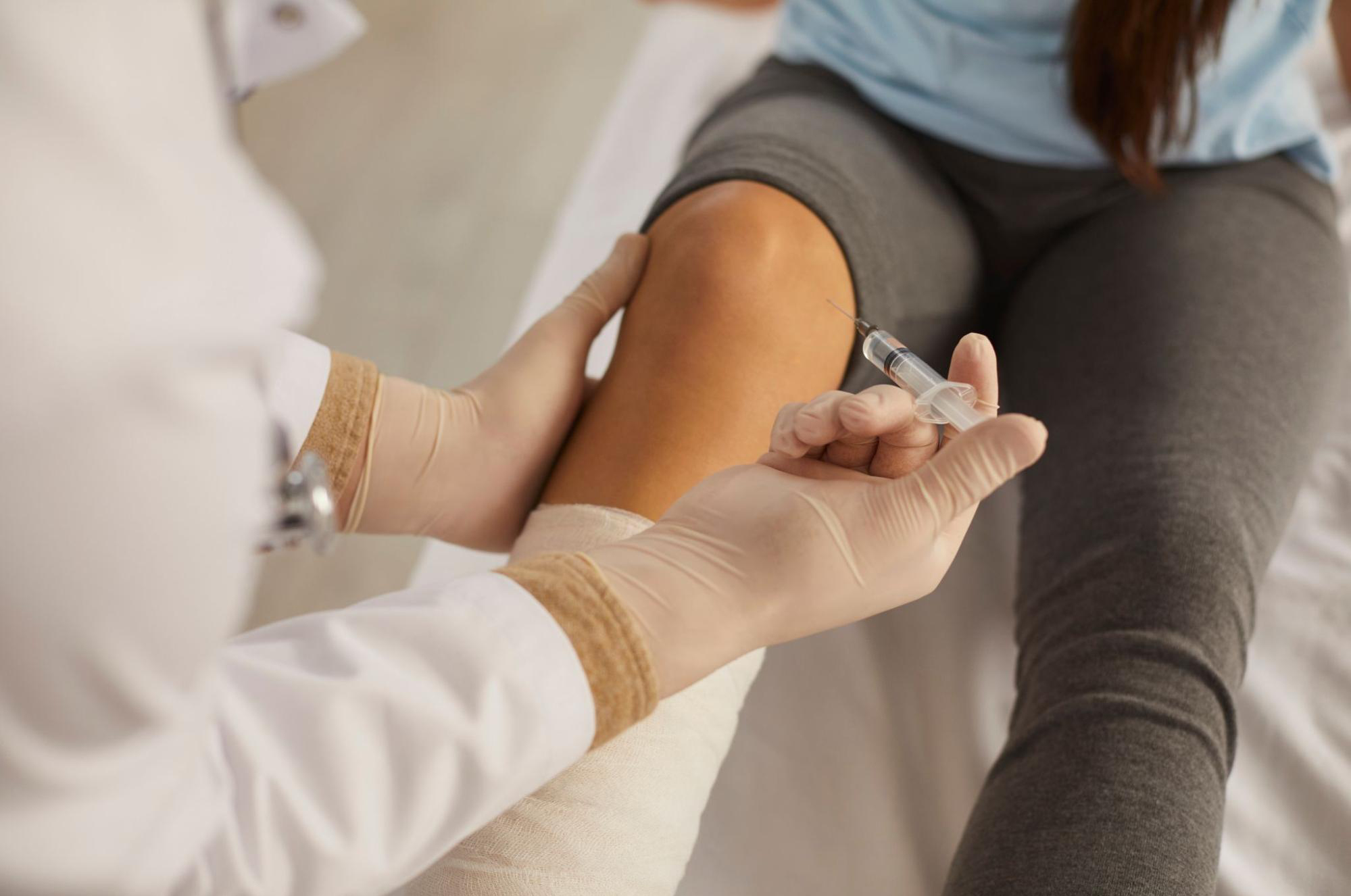
Internal Snapping Hip (Iliopsoas Tendon Snapping): Why the Groin Pops on Walking or Stair Climbing, and Peri-Articular Injection of Stem Cell Conditioned Media as a Conservative Option — A Review by Dr. Moriwaki2026.07.27
“The moment I start walking, or when I lift my leg from a lying position, I hear a ‘pop’ or ‘click’ at the front of my hip.” — This kind of snapping symptom is often observed in younger sports enthusiasts, dancers, and yoga practitioners. It usually begins painlessly, but as the repeated friction becomes chronic with every movement, it can develop into deep groin pain or discomfort. This condition is called internal snapping hip (iliopsoas tendon snapping). Dismissing it as merely “a habit of the joint clicking” carries the risk of overlooking early hip osteoarthritis or femoroacetabular impingement (FAI), and the order of differential diagnosis directly shapes the treatment strategy. In this article, Dr. Moriwaki, who oversees regenerative medicine at AVAN TOKYO Ginza, reviews the anatomical background of internal snapping hip and outlines peri-articular injection of stem cell conditioned media as a conservative option.
Key Points of This Article
・Internal snapping hip is a snapping phenomenon that occurs when the iliopsoas tendon crosses the iliopectineal eminence or the front of the femoral head during hip flexion and extension, and is common in younger populations and athletes
・Because there is often a painless period where “only the sound is present,” it tends to be left untreated, yet repeated friction can lead to chronic peri-tendinous bursitis and tendon degeneration
・It differs pathologically from greater trochanteric pain syndrome (external snapping hip), hip osteoarthritis, FAI, and groin pain syndrome, and each requires a distinct treatment design
・The foundation of conservative therapy is stretching and exercise for the iliopsoas, rectus femoris, and hip stabilizers; peri-articular injection of stem cell conditioned media is positioned as a complementary approach to the inflammatory environment
・Injection is not the first-line choice for every case of internal snapping hip; a careful judgment of indications and limitations is essential
What Is Internal Snapping Hip?
Snapping hip syndrome refers to a group of conditions accompanied by an audible snap or catching sensation during hip motion, and is anatomically classified into three types. The external type involves snapping of the iliotibial band or gluteus maximus aponeurosis over the greater trochanter; the intra-articular type is caused by labral tears or loose bodies; and this condition — the internal type — involves snapping of the iliopsoas tendon at the anterior pelvis.
The protagonist here is the iliopsoas tendon, formed by the confluence of the iliacus and psoas major. Along its course to the lesser trochanter of the femur, this tendon passes over the iliopectineal eminence — part of the superior pubic ramus — and over the anterior joint capsule of the femoral head. As the hip returns from flexion to extension, or from external to internal rotation, the moment the tendon slips medially over the edge of the bony prominence produces the characteristic snapping sound.
From “Painless Snapping” to “Pain with Inflammation”
Because it is initially painless and merely audible, many patients leave it alone for years as a “clicking habit.” However, when hundreds of snaps are repeated per day during daily activities or sports, chronic inflammation can develop in the iliopsoas bursa between the tendon and the bony prominence. Fluid accumulation in the bursa, tendon thickening, and micro-degeneration around the insertion accumulate, developing into deep groin pain or pain aggravated by hip flexion.
Conditions Commonly Confused with Internal Snapping Hip
The complaint of “pain in the front of the hip” overlaps with many conditions. Neglecting the following differential diagnoses can misalign the entire treatment design.
Differentiation from Early Hip Osteoarthritis
Hip osteoarthritis is an “intra-articular” disease centered on cartilage wear and osteophyte formation. Pain is centered at the joint space, and range-of-motion restriction — especially internal rotation — appears early. In contrast, this condition is a “peri-articular” problem of the tendon and bursa, without prominent internal rotation restriction, and is characterized by snapping sound and local tenderness. Plain X-rays for joint space narrowing and osteophytes, along with MRI evaluation of cartilage and labrum, form the basis of differentiation.
Differentiation from FAI (Femoroacetabular Impingement)
FAI is a condition where bony morphological abnormalities at the femoral head-neck junction and acetabular rim cause the labrum or cartilage to be pinched during hip flexion and internal rotation. It overlaps with this condition in that deep groin pain is provoked by forward bending or squatting, but in FAI the snapping sound is not obligatory; a positive FADIR test and increased alpha angle on X-ray serve as diagnostic clues. Coexistence is possible, making imaging-based differentiation indispensable.
Differentiation from Greater Trochanteric Pain Syndrome (External Snapping Hip)
Greater trochanteric pain syndrome is primarily caused by tendon insertion disorders of the gluteus medius and minimus, with pain located on the “outer” aspect of the hip at the greater trochanter. This condition presents with pain on the “anterior/groin” side, so the pain location itself is different. Nevertheless, coexistence is not rare, and careful history-taking and palpation of tender points are important.
Conservative Therapy — Stretching and Exercise as the Foundation
Treatment of this condition begins in principle with conservative therapy. Since tightness of the iliopsoas and rectus femoris often amplifies the snapping phenomenon, stretching these muscle groups is the first step. Additionally, stabilization training of the deep hip muscles (six external rotators) and core muscles (transversus abdominis, multifidus) reduces overload on the iliopsoas. In dancers and athletes, form correction based on motion analysis is essential for preventing recurrence. For information on joint diseases, please also refer to the guidelines of the Japanese Orthopaedic Association.
When symptoms do not improve after several months of conservative therapy, or when daily life and sports activities continue to be affected, local injection therapy is considered as the next step. Traditionally, a combined injection of local anesthetic and steroid has been used, but many patients seek alternatives out of concern about the effects of repeated steroid administration on tendon tissue.
Peri-Articular Injection of Stem Cell Conditioned Media as an Option
Stem cell conditioned media is a biological preparation containing diverse growth factors, cytokines, and exosomes secreted into the supernatant when mesenchymal stem cells are cultured. Factors such as TGF-β, IGF-1, FGF, VEGF, and HGF are thought to act on the peri-articular inflammatory cycle and contribute to establishing an anti-inflammatory, tissue-repair environment.
For this condition, ultrasound-guided local administration of stem cell conditioned media to the peri-iliopsoas tendon area and iliopsoas bursa aims to correct the chronic inflammatory environment. While steroid injections have the property of “strongly suppressing inflammation but being difficult to repeat,” stem cell conditioned media takes the directional approach of “promoting a tissue-repair environment,” so the goal axis differs. For details, please also refer to our page on joint injection with stem cell conditioned media.
Honestly Delineating Indications and Limitations
Peri-articular injection of stem cell conditioned media is not a panacea, and the following points must be made clear. First, in cases where the primary cause is pelvic morphological abnormalities or advanced organic changes in the iliopsoas tendon, injection alone does not necessarily eliminate the snapping phenomenon itself. Second, combined use with stretching and exercise therapy is a prerequisite; treating injection as complete on its own is inappropriate. Third, administration is avoided in active infection, poorly controlled systemic disease, or pregnancy. Fourth, effects vary by individual and may require multiple administrations rather than being complete in a single session. It does not guarantee efficacy and should be positioned as part of conservative therapy.
Frequently Asked Questions
Q. Is treatment necessary for painless internal snapping hip where only the sound is present?
If there are no symptoms and no interference with daily life, immediate medical intervention is not necessarily required. However, in high-intensity athletes or dancers, painless snapping repeated over months or years can progress to peri-tendinous bursitis. Preventive stretching and review of movement form are recommended.
Q. Does peri-articular injection of stem cell conditioned media eliminate the snapping sound itself?
The snapping sound itself is a phenomenon caused by physical friction between the iliopsoas tendon and the bony prominence, and injection does not change the anatomical relationship. The goal of injection is not to eliminate the snapping sound but to correct the chronic inflammatory environment resulting from repeated friction and to reduce groin pain.
Q. Should I choose steroid injection or peri-articular injection of stem cell conditioned media?
The two have different mechanisms of action. Steroids strongly suppress acute-phase pain via powerful anti-inflammatory action in the short term, but repeated administration raises concerns about effects on tendon tissue. Stem cell conditioned media targets a tissue-repair environment and is considered as an option for chronic cases or those wishing to avoid repeated steroid use. Please discuss during consultation.
Q. What if conservative therapy and injection do not lead to improvement?
In refractory cases that do not respond to long-term conservative therapy and injection treatment, surgical options such as partial tenotomy of the iliopsoas tendon may be considered. However, surgery is a last resort, and the general practice is to first go through sufficient conservative therapy and regenerative medicine approaches before deciding.
Q. What can be done in daily life to prevent recurrence?
Maintaining flexibility of the iliopsoas and rectus femoris, coordinated stabilization training of deep hip muscles and the core, and correction of the causative sports/daily movement form are the three pillars. Avoiding sudden increases in activity and not neglecting cool-down after sports involving repeated hip flexion are also important.
──────────────
Supervising Physician: Shin Moriwaki, MD
Member, Japan Society of Aesthetic Surgery (JSAS) / Member, American Academy of Aesthetic Medicine
ECFMG Certificate
──────────────
📍AVAN TOKYO 銀座 関節再生医療
AVAN TOKYO Ginza Joint Regenerative Medicine
English / 中文 / Tiếng Việt available
Inquiries via DM / LINE / Website / Phone.