


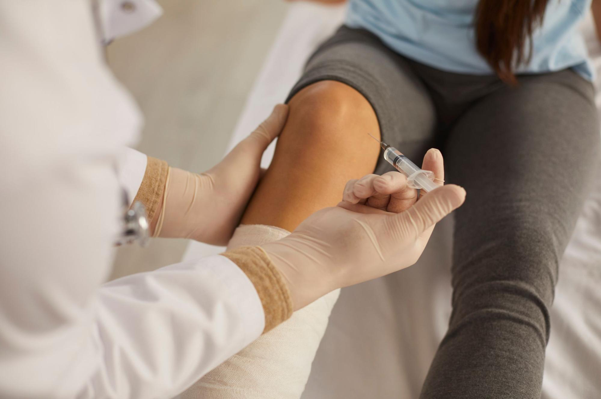
Where Should the Needle Enter for a Stem Cell Conditioned Media Knee Injection? Dr. Moriwaki Reviews Four Approaches to Knee Joint Puncture — Superolateral, Superomedial, Infrapatellar, and Medial Joint Line — and How to Improve Intra-articular Delivery2026.07.26
A “knee joint injection” for knee pain is not as simple as pressing a needle through the skin and trusting the fluid will reach the joint cavity. The knee joint space is a three-dimensional pouch bounded by the patella, femur, tibia, joint capsule, and synovial folds. The site of entry, the angle of the needle, and the depth to which it is advanced together determine both the probability that fluid actually reaches the intra-articular space and the risk of injuring adjacent tissues. When the injectate is a stem cell conditioned media knee injection — a liquid biological product containing growth factors and cytokines meant to modulate the intra-articular inflammatory environment — accurate knee joint puncture becomes the foundation on which treatment effect rests. This article reviews four commonly used approaches (superolateral, superomedial, infrapatellar, medial joint line) and the design of knee joint puncture including ultrasound-guided delivery, centered on knee osteoarthritis.
Key Points of This Article
・Delivery rates in knee joint puncture differ by approach, and landmark-only technique still leaves a nontrivial share of injections outside the joint cavity.
・The superolateral approach is easy to use in knees with flexion contracture or effusion; the superomedial approach tends to be favorable in slender patients with little subcutaneous fat.
・The infrapatellar approach is convenient in the seated position but requires careful angling to avoid the fat pad, anterior meniscal margin, and articular cartilage surface.
・The medial joint line is “easy to palpate and easy to inject,” but the needle often ends up in periarticular soft tissue rather than the joint cavity, making it a poor first choice when intra-articular delivery is the goal.
・Ultrasound-guided delivery visualizes small effusions and the location of the joint capsule, raising both the delivery rate and the safety of knee joint puncture.
Why the Choice of Approach Determines the Treatment Effect
With knee joint injections, outcomes depend not only on the nature of the fluid injected but on how accurately it lands inside the joint. When the goal is aspiration or drainage, misplacement is obvious; but with analgesics or liquid products like stem cell conditioned media, a needle that has entered subcutaneous fat or a periarticular bursa can feel deceptively normal on plunger resistance, and the misdelivery may only be recognized a week later when the patient reports “it didn’t work.” A missed knee joint puncture not only lowers efficacy but can cause swelling or discomfort from deposition into subcutaneous fat or the prepatellar bursa.
The knee joint space is a continuous cavity comprising the suprapatellar pouch above the patella, the anterior compartment bounded below by the infrapatellar fat pad, and the medial and lateral patellofemoral spaces. When effusion is abundant, most routes reach the cavity easily; but in a dry knee with minimal fluid, the capsule collapses inward, and even a small deviation lands the needle outside the joint. This is why the route must be selected in light of the individual knee — presence of effusion, body habitus, contracture, and osteophyte prominence.
Four Approaches to Knee Joint Puncture
Superolateral Approach
With the knee in extension, the needle enters just above the superolateral pole of the patella and is advanced horizontally, slightly downward and medial, into the suprapatellar pouch. This route is reported to have the highest delivery rate in knees with effusion, and it is workable even in patients with flexion contracture who cannot bend the knee. Because the suprapatellar pouch communicates with the joint cavity, injectate spreads intra-articularly. In knees with prominent osteophytes or tight lateral retinaculum, the angle must be adjusted carefully.
Superomedial Approach
The mirror image of the superolateral route, entering just above the superomedial pole of the patella and advancing laterally and slightly downward. In slender patients with thin subcutaneous fat, the patellar contour is easy to define and a stable knee joint puncture becomes feasible. However, this side lies close to the territory of the infrapatellar branch of the saphenous nerve, and some patients report cutaneous numbness, so the entry point should be chosen with care.
Infrapatellar Approach
With the knee flexed 90° in the seated position, the needle is inserted just medial or lateral to the patellar tendon and advanced slightly above the joint line. It is convenient to perform with the patient in the exam chair and tends to be low-stress for the patient. However, the infrapatellar fat pad (Hoffa’s fat pad), the anterior margin of the meniscus, and the articular cartilage surface are all nearby, so advancing too deeply risks cartilage injury. Angle and depth control matter.
Medial Joint Line Approach
With the knee slightly flexed, the needle is advanced horizontally from the medial tibiofemoral joint line. This route is commonly used for local injection to the medial meniscus, medial collateral ligament, or the pes anserinus region. It looks beginner-friendly because the joint line is easy to palpate, but in practice the needle often lodges in the periarticular soft tissues rather than the joint cavity, which makes it a poor first choice when the aim is a true intra-articular knee joint puncture. Its proper role is as a targeted periarticular injection (MCL attachment, pes anserinus).
Ultrasound-Guided Delivery vs. Landmark Technique
Landmark-only (palpation) technique for knee joint puncture depends heavily on operator experience, and in dry knees with little effusion a meaningful share of injections still miss the intra-articular target. Ultrasound guidance visualizes the joint capsule, suprapatellar pouch, fat pad, and osteophytes, and lets the operator follow the needle in real time into the cavity. For an injectate like stem cell conditioned media that aims to act on the intra-articular inflammatory and synovial environment, reliable intra-articular delivery is a precondition of treatment. General information on joint conditions is also available from the Japanese Orthopaedic Association.
Ultrasound is not mandatory in every case, but it is worth considering when: the patient has experienced ineffective past injections, obesity makes bony landmarks hard to palpate, there is severe flexion contracture, or the knee is essentially dry without effusion. For more on our stem cell conditioned media joint injections, see the joint injection page.
Aftercare Following Knee Joint Puncture
Heaviness or discomfort at the injection site may occur for a day to a few days after the puncture. This is a tissue reaction to the needle itself and usually resolves on its own. On the day of injection, avoid strenuous exercise, long hot baths, and heavy alcohol intake; from the next day, resume normal activities and pursue the physician-directed strengthening of periarticular muscles in parallel. If swelling rapidly worsens, if fever develops, or if severe pain that prevents joint motion persists for several days, other complications such as infection must be considered, and prompt consultation is essential.
A stem cell conditioned media knee injection is not a one-shot treatment that regrows cartilage but a treatment that acts stepwise on the intra-articular inflammatory and synovial environment. Improving the delivery rate of knee joint puncture corresponds to setting up the starting line properly. Individual variation and limits of indication are inevitable, and response varies with the stage of osteoarthritis, extent of cartilage loss, body weight, and lifestyle.
Frequently Asked Questions
Q. How painful is a knee joint injection?
There is a brief prick on entry and a sense of pressure as the fluid enters. Topical anesthesia and a fine needle can reduce discomfort, but pain perception varies from person to person.
Q. Can the patient choose which route the needle uses?
You can share your preferences, but the final choice of knee joint puncture route is made by the physician based on the amount of effusion, body habitus, degree of contracture, and location of osteophytes.
Q. Is ultrasound guidance always necessary?
Not in every case, but it is recommended when the knee is dry with little effusion, when past injections failed to yield relief, or when palpation is difficult in an obese patient — to ensure intra-articular delivery.
Q. Does stem cell conditioned media regenerate cartilage?
Conditioned media is not a treatment that directly “regrows” cartilage; it aims to modulate the intra-articular inflammatory and synovial environment to improve pain and function. Indications and limits vary by individual, and response depends on the stage of osteoarthritis.
Q. What intervals between sessions are recommended?
An initial loading phase typically involves several sessions at intervals of a few weeks, transitioning to a longer maintenance interval based on response. Details will be discussed at your consultation.
──────────────
【Supervising Physician】Shin Moriwaki, MD
Member, Japan Society of Aesthetic Surgery (JSAS) / American Academy of Aesthetic Medicine
ECFMG Certificate
──────────────
📍AVAN TOKYO Ginza Joint Regenerative Medicine
English / 中文 / Tiếng Việt available
Inquiries welcomed via DM / LINE / Website / Phone.