


Sharp, pinpoint pain just in front of and below the inner ankle bone: distinguishing painful accessory navicular syndrome from lingering sprain pain and flatfoot pain — the chronic micro-injury cycle at the accessory bone on the medial navicular and the posterior tibial tendon insertion in adolescent girls and amateur runners, and where a stem cell conditioned media peri-ankle injection can help by calming the inflammatory environment at the tendon insertion and peri-articular bursa versus where it cannot physically remove the accessory bone or rebuild the medial longitudinal arch — organized by Dr. Moriwaki2026.08.15
After exercise or a long day of walking, when you press the spot just in front of and slightly below the medial malleolus with your thumb, a sharp, pinpoint ache flares — this is often ‘painful accessory navicular,’ a chronic pain seen frequently in adolescent girls, amateur runners, and soccer or volleyball players. It arises at the boundary between an accessory bone (os tibiale externum) on the medial edge of the navicular and the insertion of the posterior tibial tendon. It is often dismissed for years as ‘leftover pain from a sprain’ or ‘the flatfoot is just like that,’ managed with patches and taping until running and even walking down stairs become difficult. Dr. Moriwaki of AVAN TOKYO Ginza Joint Regenerative Medicine walks through the pathology of painful accessory navicular, its differential diagnosis, and where a stem cell conditioned media peri-ankle injection can reach — and where it cannot — while placing it alongside orthotics, exercise therapy, medication, and surgery.
Key points of this article
・Painful accessory navicular is fundamentally a chronic cycle of micro-injury and failed repair at the accessory bone on the medial navicular and the posterior tibial tendon insertion
・Mistaking it for lingering sprain pain or generic flatfoot pain causes patients to miss the correct entry point to orthotic design, exercise therapy, and injection options
・Stem cell conditioned media peri-ankle injection is a conservative option aimed at calming the inflammatory environment at the tendon insertion and peri-articular bursa
・Physical removal of the accessory bone itself and structural rebuilding of the arch cannot be achieved by injection; surgical excision is considered for refractory cases
・Response and limits vary considerably between individuals, and it is essential to agree on a combination plan with orthotics, exercise therapy, and medication, and to set the timing of response assessment, at the very first visit
What is painful accessory navicular — what is actually happening at the accessory bone and the posterior tibial tendon insertion
A ‘second bone’ on the medial side of the navicular
This condition refers to the pain that arises in people who congenitally have an accessory bone, the ‘os tibiale externum,’ on the medial edge of the navicular. This bone is an anatomical variant present in roughly 10–20% of the general population. Many people never experience symptoms in a lifetime, while a certain proportion begin to feel pain from adolescence onward. In particular, when a larger accessory bone (Veitch type II) sits right where the posterior tibial tendon inserts into the navicular, the traction stress from the tendon concentrates at this area and is more likely to cause pain.
Micro-injury repeated each time the posterior tibial tendon pulls
The posterior tibialis is a key muscle that supports the medial longitudinal arch and stabilizes the foot during walking, running, and jumping landings. When its tendon inserts across the boundary between the navicular and the accessory bone, every single step applies micro-avulsion stress between the tendon and the accessory bone. When adolescent athletics, dance, or long-distance running concentrate load on this area, a chronic cycle of inflammation and failed repair begins at this boundary, the fibrocartilaginous connection between the accessory bone and the navicular gradually becomes unstable, and pain becomes fixed. Understanding that this pain is not merely a lingering echo of a bruise or sprain — but rather an entheseopathy at the tendon insertion combined with chronic micro-injury of the fibrocartilage connection — is the starting point for treatment design.
Why it is easily misdiagnosed — differentiating from post-sprain sequelae, flatfoot pain, and posterior tibial tendon dysfunction
How it differs from the sequelae of an ankle sprain
Most patients arrive saying, ‘The pain never went away after I twisted my ankle.’ But careful palpation of the pain center reveals a single pinpoint tenderness not at the anterior talofibular ligament insertion on the outer side, but 2–3 cm in front of and below the medial malleolus, right at the medial edge of the navicular. In true sprain sequelae, the typical residual findings are a sense of instability and lateral tenderness even after weight-bearing pain improves. In this condition, we repeatedly confirm the finding, ‘Press only that one spot on the medial side and the patient jumps.’ When this pinpoint tenderness is combined with ‘pain at the same spot when the posterior tibial tendon contracts against resistance’ (resisted inversion pain), diagnostic accuracy improves substantially.
Continuity with flatfoot pain and posterior tibial tendon dysfunction (PTTD)
This condition and flatfoot exist on a continuum: in some people, the presence of the accessory bone reduces the mechanical efficiency of the posterior tibial tendon, and the medial longitudinal arch gradually collapses. Conversely, in the early stages of acquired posterior tibial tendon dysfunction (PTTD), pain around the navicular insertion can appear in a form easily confused with this condition. Useful functional evaluations for distinguishing the two include whether the patient can perform a single-leg heel raise (this progressively becomes impossible in PTTD) and whether hindfoot valgus deformity is progressing. Which entry point — orthotics, exercise therapy, injection, or surgery — you choose depends entirely on the resolution of this differential diagnosis. General information on joint disorders is also available on the website of the Japanese Orthopaedic Association.
What a stem cell conditioned media peri-ankle injection can and cannot target
What it can target: the inflammatory environment at the tendon insertion and surrounding bursa
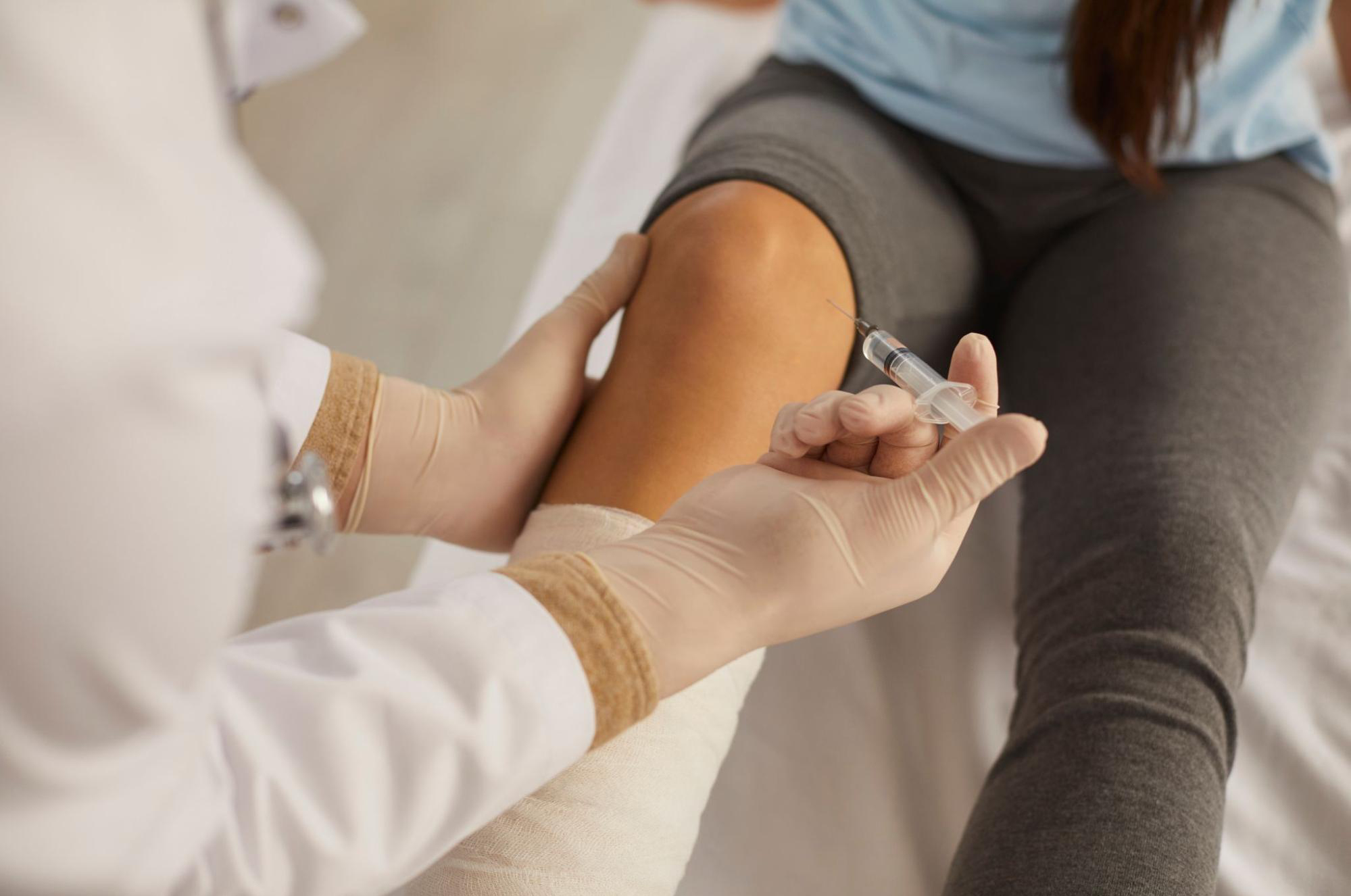
Stem cell conditioned media contains growth factors such as TGF-β, IGF-1, FGF, and VEGF, along with regulatory signaling molecules carried in extracellular vesicles (exosomes). It is expected to work on the local environment of a tendon insertion trapped in a cycle of chronic inflammation and failed repair. For this condition, the peri-ankle injection is organized to address three layers with separate targets — around the junction of the accessory bone and the navicular, the insertion of the posterior tibial tendon, and the bursa immediately deep to it — carefully delivered under ultrasound guidance. What it targets is strictly the ‘inflammatory and repair environment,’ and unlike analgesics or steroids, it is not an injection designed to directly block pain sensation. See also this page for details on stem cell conditioned media joint injections.
What it cannot target: physical removal of the accessory bone and rebuilding of arch architecture
At the same time, we need to be clear at the very first visit about what injection ‘cannot do.’ The accessory bone itself will not shrink, fuse with the navicular, or disappear because of an injection. Similarly, the length of the posterior tibial tendon or the shape of the medial longitudinal arch cannot be structurally redesigned by injection alone. Those roles belong to orthotics (insoles that support the medial longitudinal arch), exercise therapy for the calf and lower leg musculature, and — for refractory cases — surgical excision of the accessory bone (procedures such as the Kidner operation). Conditioned media peri-ankle injection is not a ‘substitute’ for any of these; the honest positioning is as a ‘complement’ that adjusts the baseline of inflammation and pain within each of these treatment lanes.
Designing treatment — placing orthotics, exercise therapy, conditioned media injection, and surgery in order
Start with orthotics and exercise therapy
Conservative treatment for painful accessory navicular is built on the foundation of rest, icing, and oral NSAIDs during flare periods, along with introducing an insole that supports the medial longitudinal arch and progressive strengthening plus stretching of the posterior tibialis and triceps surae. Off-the-shelf orthotics can be effective, but for younger athletes, custom-made insoles based on foot-shape evaluation are often useful, and it is critical that the insole design does not press directly on the accessory bone. Simply pausing running and jumping in the short term and switching to low-impact aerobic exercise (water walking, cycling) is not uncommonly enough to reduce pain within several weeks.
Where to place a stem cell conditioned media peri-ankle injection
We consider conditioned media peri-ankle injection alongside orthotics and exercise therapy for chronic cases where pain recurs despite several months of conservative therapy, or for younger athletes who need to keep competing through a season. Injections are typically arranged as a series of 2–3 sessions at intervals of several weeks to several months, and response is assessed with objective indicators such as VAS pain, tenderness intensity, and single-leg standing stability. When the response is poor, it is worth stepping back to reassess the diagnosis — progression to PTTD, coexistence of other enthesopathies, or reconsideration of the Veitch classification.
Indications for surgery (excision of the accessory bone)
When several months of the above conservative therapy and peri-ankle injection still leave pain that disrupts daily life, excision of the accessory bone (Kidner procedure) — or, depending on the case, a procedure including re-attachment of the posterior tibial tendon — is considered. The surgical decision is in the domain of the orthopedic surgeon, and the injections at our clinic are strictly positioned within conservative management. We cannot assert that ‘trying hard with injections will always avoid surgery’; the most important thing is to avoid missing the timing of surgery based on the degree of progression and the patient’s life circumstances.
Frequently asked questions
Q. Can painful accessory navicular appear for the first time in adulthood?
Yes, it can. The accessory bone itself is a congenital anatomical variant, but while the most common first onset is triggered by the athletic loads of adolescence, we clinically often see cases that first develop in the 40s or 50s, triggered by a sudden start of long-distance walking or mountaineering as an adult, or by changes in foot loading after weight gain, pregnancy, or childbirth. Do not rule out this condition based on age alone.
Q. How many stem cell conditioned media peri-ankle injections are needed before response can be judged?
Typically, we set a series of 2–3 sessions at intervals of several weeks to several months, and reassess VAS pain, tenderness intensity, and functional limitation 1–3 months after the series ends. Response varies considerably between individuals — from patients who feel clear changes after a single injection, to those who see little improvement even after completing a series. When response is poor, we recommend either revising the orthotic and exercise plan, or stepping back to the differential diagnosis.
Q. How do we decide whether to do surgery (excision of the accessory bone) first, or persist with injection?
We judge it based on how much and where the pain disrupts daily life, work, and sports; how well it responds to orthotics and exercise therapy; and the size and Veitch type of the accessory bone. The academic or sports season schedule and the social feasibility of rest are also factors. The decision about surgery is made by the orthopedic surgeon, and conditioned media injection is positioned within the conservative management lane.
Q. Can I continue sports activities during treatment?
During flare periods, we strongly recommend pausing running and jumping in the short term and switching to low-impact aerobic exercise. After a conditioned media injection, we set a downtime of several days to a week, and gradually return to competition load while supporting the medial longitudinal arch with taping or orthotics. Pushing through pain while continuing sports risks progression to posterior tibial tendon dysfunction and contralateral compensation-related knee and hip disorders.
Q. Are orthotics (insoles) really necessary?
In treating this condition, orthotics are not ‘a tool for enduring pain’ but a pillar of treatment that disperses the traction stress on the posterior tibial tendon insertion and avoids direct pressure on the accessory bone. If injection is repeated without orthotics, the mechanical stress source remains, and symptoms are more likely to recur. Building treatment as a three-piece set of orthotics, exercise therapy, and injection is the basic approach.
──────────────
Supervising physician: Shin Moriwaki, MD
Member, Japan Society of Aesthetic Surgery (JSAS) / Member, American Academy of Aesthetic Medicine
ECFMG Certificate
──────────────
📍AVAN TOKYO 銀座 関節再生医療
AVAN TOKYO Ginza Joint Regenerative Medicine
English / 中文 / Tiếng Việt supported
Please contact us via DM / LINE / Website / Phone.