


Sudden Adult-Onset Flatfoot May Be Posterior Tibial Tendon Dysfunction — Where Stem Cell Conditioned Media Peritendinous Injection May Help and Where It Cannot, Organized by Dr. Moriwaki2026.07.23
You get out of bed in the morning, take your first steps, and feel a heavy ache along the arch of your foot. You used to walk without any problem, yet at some point the medial arch has collapsed, your foot has become flat, and the inner side of your ankle throbs after long walks. Behind symptoms like these, we frequently find a tendon disorder called posterior tibial tendon dysfunction. Posterior tibial tendon dysfunction (PTTD) is a condition in which the main tendon supporting the medial longitudinal arch undergoes degeneration and loses function. Left untreated, it progresses into adult-acquired flatfoot and eventually leads to deformity of the subtalar and ankle joints. Recently, peritendinous injection of stem cell conditioned media has emerged as one option to complement conservative care, but the line between where it may help and where it cannot must be drawn carefully. In this article, from the perspective of Dr. Moriwaki, we organize the pathology of posterior tibial tendon dysfunction and honestly map out where such injection can and cannot contribute, alongside orthotics, exercise therapy, and surgical options.
Key Points of This Article
・Posterior tibial tendon dysfunction is a leading cause of adult-acquired flatfoot, most frequently seen in women aged 40 to 60
・Degeneration and partial tearing of the tendon supporting the medial longitudinal arch is the underlying mechanism, and progression leads to ankle and subtalar deformity
・Orthotics and exercise therapy remain the foundation; from Johnson stage III onward, surgical intervention is considered
・Peritendinous injection of stem cell conditioned media is positioned as a conservative complement that acts on the inflammatory and reparative environment around the degenerated tendon
・Response varies widely between individuals; when the tendon is largely torn or bony deformity has set in, injection alone cannot be expected to bring improvement
What Is Posterior Tibial Tendon Dysfunction
The tibialis posterior is a deep muscle of the posterior calf. Its tendon passes behind the medial malleolus and attaches to the navicular and to the bases of the metatarsals. This tendon plays a central role in lifting the medial longitudinal arch during gait and in generating push-off. When the tendon degenerates and elongates, pain and swelling first appear just below and behind the medial malleolus, the arch progressively collapses, and the characteristic “too many toes sign” becomes visible from behind. Difficulty performing a single-leg heel rise is another representative sign of this condition.
A Leading Cause of Adult-Acquired Flatfoot
While most pediatric flatfoot is skeletal and asymptomatic, gradual flattening that appears in adulthood often has this tendon disorder behind it. Age-related tendon degeneration, chronic overload, obesity, hormonal changes in women, diabetes, and systemic conditions such as rheumatoid arthritis are thought to combine and precipitate onset. The higher incidence in middle-aged and older women is often attributed to the interaction between the post-menopausal hormonal environment and collagen metabolism in tendon tissue.
The Johnson Classification and Natural Course
The Johnson classification (stages I to IV) is widely used. Stage I involves tendon inflammation with preserved function; stage II shows tendon elongation and flatfoot deformity with retained flexibility; stage III is characterized by fixed deformity with bony changes; and stage IV extends to valgus deformity of the ankle joint. As stages advance, the benefits of conservative therapy become more limited, and from stage III onward, procedures such as subtalar or triple arthrodesis are considered. For general information on joint and foot disorders, please also refer to publicly available resources such as the Japanese Orthopaedic Association.
What Can Stem Cell Conditioned Media Peritendinous Injection Offer for Posterior Tibial Tendon Dysfunction
Treatment of this disease begins with orthotics (medical arch-support insoles, UCBL braces, and AFO when needed) and exercise therapy (eccentric strengthening of the tibialis posterior, calf stretching). Short-term anti-inflammatory medication may be used in acute phases, but repeated steroid injections into the tendon carry a risk of tendon weakening and require caution. Hyaluronic acid injection is not considered a standard option for this condition either. It is in this gap that peritendinous injection of stem cell conditioned media enters the picture as “another conservative complement.”
Adjusting the Tissue Repair Environment
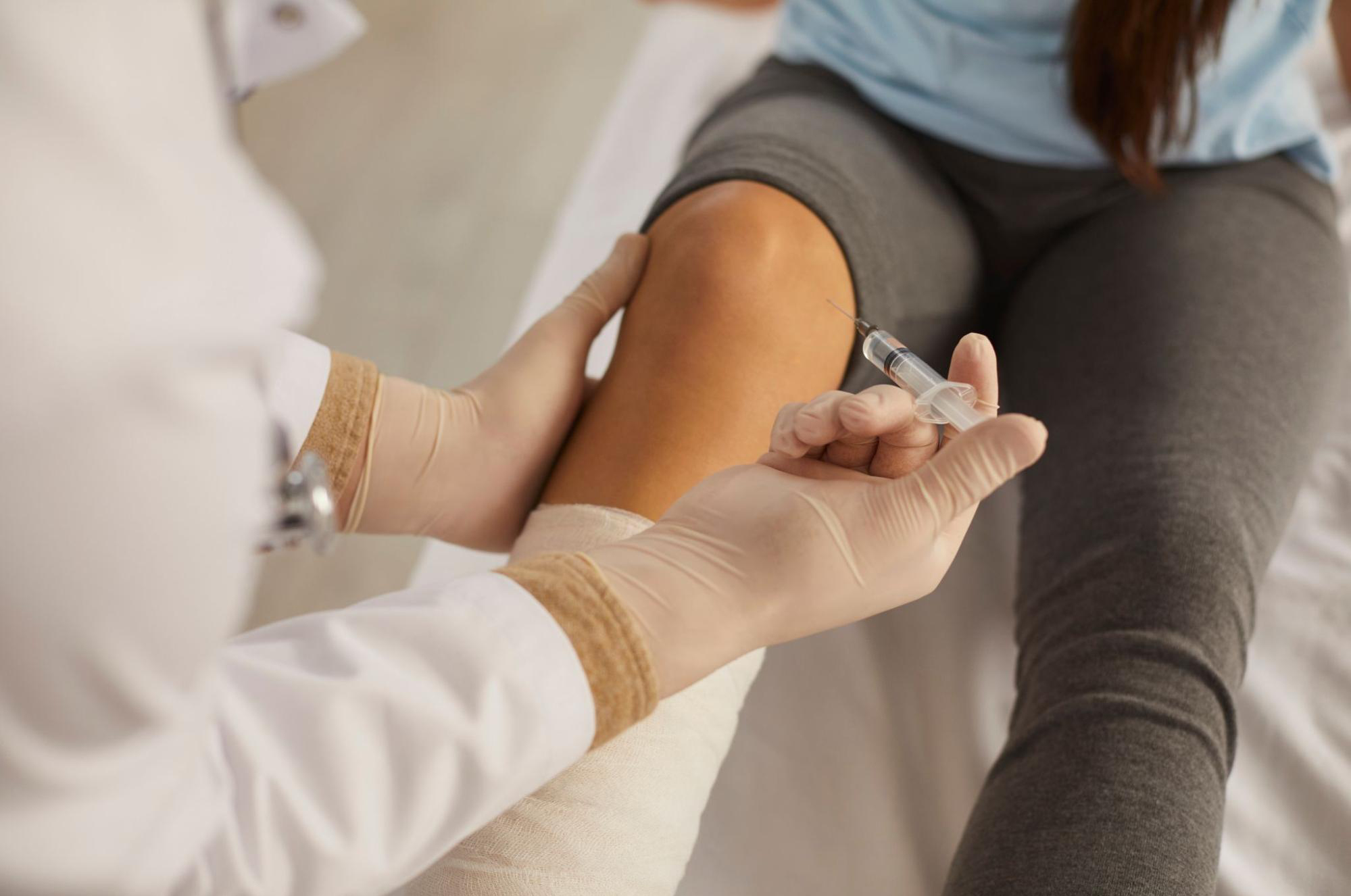
Stem cell conditioned media is reported to contain multiple growth factors (TGF-β, IGF-1, FGF, VEGF), anti-inflammatory cytokines, and exosomes. Tendon tissue is inherently poor in blood supply, and repair at a degenerated tendon insertion is slow — one reason these conditions become chronic. Peritendinous injection of stem cell conditioned media aims to act on this “difficult-to-heal environment,” calming the inflammatory cycle and supporting the repair environment around the tendon. However, this is not a treatment that “shortens an elongated tendon” or “re-attaches a torn tendon.” It is a complementary approach that modulates the biological environment around the tendon, and this framing should be shared from the outset.
Indication and Limitations by Stage
This injection may play a meaningful role as conservative complement primarily in Johnson stage I to early stage II, where tendon inflammation and partial degeneration are present but deformity remains flexible and orthotics and exercise therapy can be combined. From stage III onward, once bony deformity has set in at the subtalar or midtarsal joints, expecting functional recovery from injection alone is unrealistic and orthopedic surgery becomes the first choice. When extensive complete tearing or fatty degeneration of the tendon is present, improvement from injection is limited. For more on treatment options directed at joints and peritendinous tissues, please also see our page on stem cell conditioned media joint injection.
Treatment Design and Combined Therapies in Practice
Orthotics and Exercise Therapy Are the Foundation
Peritendinous injection for this condition is never designed as a standalone treatment; it is always paired with orthotics and exercise therapy. Support of the longitudinal arch by medical arch-support insoles or UCBL braces reduces load on the tibialis posterior and provides the “resting environment” required for tendon repair — a role that is genuinely indispensable. Standard exercise components include eccentric strengthening of the tibialis posterior with a resistance band (toe-in movements), stretching of the gastrocnemius, soleus, and Achilles tendon, and single-leg balance training. The misconception that “an injection removes the need for exercise” is precisely what undermines outcomes most.
Assessing Response and Session Design
After injection, we typically evaluate over several weeks to several months, using objective indicators such as pain scores (VAS/NRS), the ability to perform a single-leg heel rise, walking distance before pain onset, and change in the medial longitudinal arch angle. When response is poor or deformity progression is confirmed, we should switch to orthopedic re-evaluation rather than continue injections passively. Rather than expecting “dramatic pain relief from one session,” a realistic framing is to see this as “raising the baseline” of a conservative program built on orthotics and exercise.
Who This Is Not For, and Who Requires Caution
Active joint or skin infection, poorly controlled diabetes or rheumatoid arthritis, stage IV disease with fixed deformity, and cases of extensive complete tendon rupture are all situations where this injection is generally not indicated or requires careful judgment. Patients on anticoagulants such as warfarin, DOACs, or antiplatelet agents need to consult their primary physician in advance regarding continuation or withdrawal. Because safety data are limited during pregnancy and lactation, timing considerations are generally recommended.
Frequently Asked Questions
Q. If I receive stem cell conditioned media injection for posterior tibial tendon dysfunction, will my flatfoot return to normal?
This is not a treatment aimed at fully restoring the original arch. It is positioned as a conservative complement intended to calm inflammation around the tendon, condition the repair environment, and slow symptom progression. Functional improvement is most reasonable to expect only in early stages before bony deformity has set in.
Q. Can I be treated with injection alone, without orthotics?
We do not recommend this. Unless the load on the tibialis posterior is reduced through orthotics and exercise therapy, the repair environment for the tendon cannot be established. This injection is designed to be combined with medical insoles and exercise therapy as a prerequisite.
Q. When can I expect to feel improvement?
Individual variation is large and we cannot make definitive claims. Because tendon repair takes time, in most cases evaluation is spread across several weeks to several months. This must be understood in advance — it is not a treatment where a single injection dramatically eliminates pain.
Q. Can I avoid surgery?
In early Johnson stage I to II, aiming to avoid surgery through the combination of orthotics, exercise therapy, and peritendinous injection is a realistic design. From stage III onward, however, with fixed deformity, we cannot promise that injection will avert surgery, and consultation with an orthopedic surgeon becomes essential.
Q. Is this covered by insurance?
Stem cell conditioned media peritendinous injection is out-of-pocket, self-pay care. Orthotics and exercise therapy are often available within insured care, so we recommend budgeting across the total of your conservative regimen rather than a single item.
──────────────
[Supervising Physician] Shin Moriwaki (Supervising Doctor)
Member, Japan Society of Aesthetic Surgery (JSAS) / Member, American Academy of Aesthetic Medicine
ECFMG Certificate
──────────────
📍AVAN TOKYO 銀座 関節再生医療
AVAN TOKYO Ginza Joint Regenerative Medicine
English / 中文 / Tiếng Việt supported
Please contact us via DM / LINE / Website / Phone.