


Should Fat Grafting Breast Nodules Be Removed? Four Medical Criteria for Calcified Lump Removal Explained by a Doctor2026.07.22
One of the most common post-operative concerns raised by patients who have undergone fat grafting to the breast is the presence of palpable nodules. Calcified lumps can also be discovered incidentally during breast cancer screening, and many patients struggle with the decision of whether to proceed with fat grafting nodule removal or to simply monitor them over time. In this article, Dr. Moriwaki of AVAN TOKYO Ginza Liposuction Clinic explains when fat grafting nodule removal should be considered, and which lumps can be safely observed, from a clinical medical perspective.
Key Points of This Article
– Most nodules after fat grafting can be safely followed up; fat grafting nodule removal is reserved for a limited set of cases
– The decision to remove is based on four criteria: size, growth trend, calcification pattern, and impact on breast cancer screening
– Oil cysts often resolve with simple needle aspiration alone
– Micro-calcifications and macro-calcifications are distinguished on mammography and managed differently
– Avoid self-diagnosis and remain under the ongoing care of a physician experienced in fat grafting
What Are Post Fat Grafting Nodules?
Nodules palpated after fat grafting are not all the same. The most common are oil cysts, capsular collections where transplanted fat cells failed to engraft, liquefied, and became encased in a thin membrane. They tend to be soft, mobile, and appear as well-defined hypoechoic or anechoic lesions on ultrasound. The next most common are fat necrosis nodules, small firm lesions where necrotic fat cells are surrounded by scar tissue; these are usually hard and poorly mobile. When calcium eventually deposits around or within the necrotic tissue, calcified nodules form, appearing white on mammography. All of these are benign changes and are not immediately dangerous. However, whether fat grafting nodule removal is truly required must be judged carefully based on the type of lump, its size, symptoms, and imaging findings.
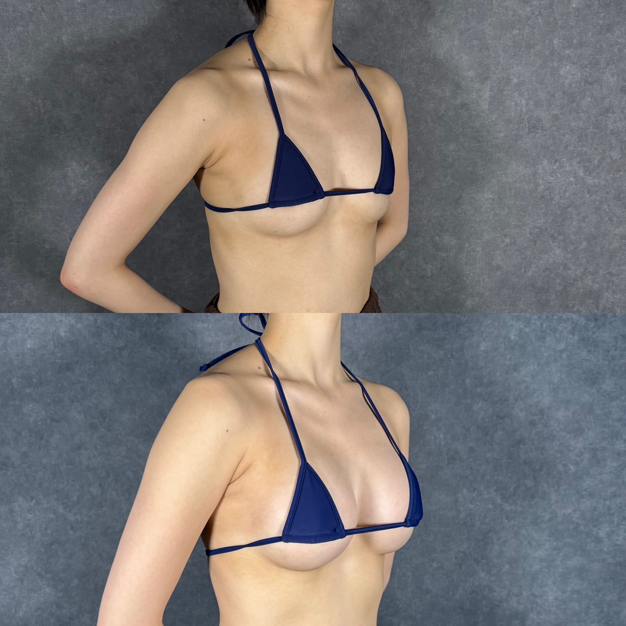
Four Criteria for Fat Grafting Nodule Removal
In clinical practice we decide how to manage a nodule based on four viewpoints. First is size: palpable firm lumps larger than 2 cm often affect quality of life and become candidates for removal. Second is growth trend: enlargement over time or progressive hardening calls for active intervention. Third is the calcification pattern on mammography. Linear, branching, or pleomorphic features suggestive of malignancy require tissue diagnosis (biopsy) even when benign etiology is likely. Fourth is impact on breast cancer screening; if the lump obscures imaging interpretation, removal may be recommended to preserve future screening accuracy. Conversely, small, stable, asymptomatic macro-calcifications rarely need surgery and can be safely monitored. These four axes guide our decisions on fat grafting nodule removal.
Classification and Management of Calcified Nodules
Calcifications after fat grafting are broadly divided on mammography by size and shape. Micro-calcifications under 1 mm in diameter can overlap with early findings of breast cancer, making differentiation difficult in fat grafting patients. In contrast, typical calcifications after fat grafting are macro-calcifications ranging from several millimeters to over 1 cm, with smooth margins that often appear eggshell-like and are classified as benign. Macro-calcifications generally require only observation, but removal is considered when patients report strong palpable discomfort or when multiple macro-calcifications cluster and cause visible contour changes. At our clinic we recommend ultrasound follow-up at 6 months, 1 year, and annually thereafter for patients who have undergone fat grafting, so any change can be detected early.
Surgical Method and Downtime
Fat grafting nodule removal varies with the depth and size of the lump. Superficial oil cysts of 1-2 cm can be treated under local anesthesia through a few-millimeter incision with needle aspiration, leaving virtually invisible scars. Firm fat necrosis nodules or those with calcifications, being non-liquefied, are more reliably excised together with the capsule. Incisions are placed along inconspicuous lines such as the areolar border or inframammary fold. Downtime typically includes 1-2 weeks of swelling, 2-3 weeks of bruising, and about 1 month of taping; vigorous exercise and upper-body gym training should be avoided for about 4 weeks. To prevent post-excision depressions, small amounts of fat re-injection or repositioning of surrounding tissue may be added. For safety standards in aesthetic surgery, please refer to the Japan Society of Aesthetic Surgery. For more detail, please also see our liposuction and fat grafting column list.
Frequently Asked Questions
Q. Do nodules after fat grafting shrink on their own?
Small oil cysts may gradually shrink as the oil content is absorbed by the body. Firm lumps caused by fat necrosis or calcification, however, rarely disappear spontaneously and usually remain roughly the same size. Regular ultrasound examinations are used to confirm that no changes are occurring.
Q. Can these nodules be mistaken for breast cancer?
Macro-calcifications after fat grafting are usually easy to identify as benign, but careful differentiation is needed when micro-calcifications are also present. Informing the screening facility in advance that you have had fat grafting, and combining ultrasound and MRI with mammography, reduces the risk of misinterpretation.
Q. Are the scars from removal surgery visible?
The incision is only a few millimeters to about 1 cm, placed along inconspicuous lines such as the areolar edge or inframammary fold, so mature scars are usually barely noticeable. Individual variation in scar hypertrophy does exist.
Q. How can nodules be prevented?
The strongest prevention is to keep each session’s injection volume within the medically acceptable range and to inject small aliquots through many points using a fine cannula (micro-injection technique). Smoking, rapid weight loss, and strong postoperative compression should also be avoided as they impair fat survival.
──────────────
[Supervising Physician] Shin Moriwaki, MD
Member of the Japan Society of Aesthetic Surgery (JSAS) / Member of the American Academy of Aesthetic Medicine
ECFMG Certificate
──────────────
📍AVAN TOKYO GINZA LIPOSUCTION CLINIC
English / 中文 / Tiếng Việt supported
Appointments and consultations are available via
DM / LINE / Website / Phone.