


Pre-Op Breast Ultrasound Before Hybrid Augmentation: Screening for Hidden Masses2026.09.11
Hybrid breast augmentation combines a silicone implant with autologous fat grafting, and its clinical quality depends on a step that starts well before the incision: a hybrid augmentation preoperative ultrasound to screen for occult breast masses. At our clinic we perform breast ultrasound both in the pre-op consultation and again in the operating room just before anesthesia induction, checking for fibroadenomas, cysts, and any lesion suspicious for malignancy. This article uses one case — a woman in her 30s who underwent circumferential upper-arm and chest liposuction as donor sites, with a subglandular 250 cc Motiva-type round implant via axillary incision and subcutaneous fat injection — to explain why hybrid augmentation preoperative ultrasound matters and how our two-step protocol works.
Key Points
・A hybrid augmentation preoperative ultrasound is a non-invasive screening step to detect fibroadenomas, cysts, and rare malignant lesions inside the breast before surgery.
・Once a silicone implant is placed subglandularly, palpation and imaging of the native gland become harder, so mapping the breast beforehand is especially important.
・We use a two-step protocol: one ultrasound at the pre-op consultation and another in the operating room just before anesthesia, so the day-of-surgery status is confirmed.
・Most findings are benign, but suspicious features (irregular margins, rich vascularity, rapid growth) mean specialized workup takes priority over cosmetic surgery.
・Ultrasound adds essentially no downtime and functions as the safety anchor for the entire hybrid augmentation plan.
Why Ultrasound Is Needed Before Hybrid Augmentation
Breasts targeted for augmentation not infrequently contain silent benign lesions such as simple cysts or fibroadenomas. These usually cause no daily symptoms, but once a silicone implant sits under the gland, palpation feels different and mammography views become harder to interpret because of overlapping shadows. In other words, it becomes difficult to tell whether a mass was already there or appeared after surgery. A hybrid augmentation preoperative ultrasound maps the gland in advance, so if the patient later feels a lump we can compare with the baseline instead of ordering redundant tests or generating unnecessary anxiety.
Ultrasound Is About More Than Size
Pre-op ultrasound is not just a size check. We look at margin clarity, internal echo pattern, posterior acoustic features, vascular signal, and the relationship to surrounding parenchyma. Most findings fall into a clearly benign category, but if we see irregular margins, hypervascularity, or a rapidly enlarging lesion, we defer the hybrid augmentation and refer for specialist evaluation. Bending clinical judgment because “the surgery is already booked” is exactly what long-term safety cannot afford.
Our Two-Step Hybrid Augmentation Preoperative Ultrasound Protocol
The first ultrasound is performed at the pre-op consultation, where we set the plan (proceed, observe, or refer). On the day of surgery we ultrasound the breast again inside the operating room, immediately before anesthesia, to catch anything that may have appeared or changed in the interval. Breast tissue can look different depending on the menstrual cycle and hormonal status, so a same-day re-check has independent value. In this case, the hybrid augmentation preoperative ultrasound was performed at both time points and confirmed “no significant mass” before the operation started.
Treatment, Cost, and Risks for This Case
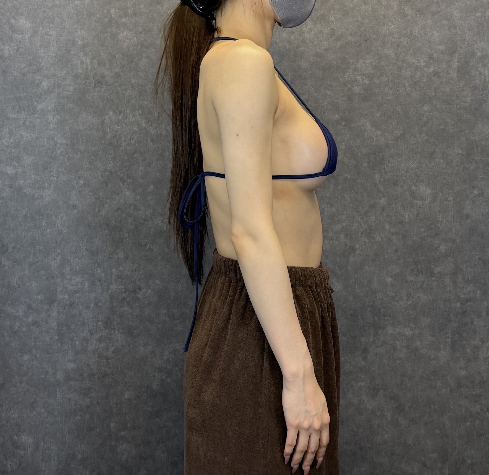
・Treatment: Circumferential upper-arm and chest subcutaneous liposuction as donor sites; a Motiva-type 250 cc round silicone implant placed subglandularly via axillary incision; centrifuged pure fat injected into the subcutaneous layer of both breasts — a hybrid augmentation.
・Cost: Please see our pricing page. Final quotations are confirmed at the in-person consultation.
・Main risks / side effects: bruising, swelling, fibrous contracture, asymmetry, infection, capsular contracture, implant malposition or rupture, fat-related nodules (oil cysts) or calcifications, and rare complications such as fat embolism.
・Results vary between individuals; photos represent one example only.

Design Rationale: Subglandular Implant + Subcutaneous Fat
In this case we placed a soft Motiva-type round 250 cc implant in the subglandular plane and limited fat injection to the subcutaneous layer. A subglandular implant tends to give a structurally stable upper pole and cleavage line, but in thin patients the implant edge or rippling can become visible. Layering centrifuged pure fat thinly into the subcutaneous plane softens that outline and improves both look and feel. Injecting fat directly into the plane in contact with the implant is avoided, both for graft survival and for safety.
Intra-Op Findings and Contracture Care
Hybrid augmentation combines pocket creation, liposuction, and fat grafting in a single continuous flow. In this case the subglandular pocket created through the axillary incision showed no significant bleeding or anatomical anomaly; fat was harvested from the arm and chest, centrifuged, and injected into the subcutaneous layer of both breasts in roughly balanced amounts. Post-operatively a bolero-type compression garment is worn to guide even fibrous remodeling of the arm during the contracture phase (roughly 1–3 months). On the breast side we balance compression and cooling individually so the graft-take phase is not disturbed by excess inflammation. For related articles, see our liposuction column index. For general aesthetic surgery safety standards, refer to the Japan Society of Aesthetic Surgery.
Frequently Asked Questions
Q. Can I use a breast ultrasound performed at another clinic instead?
Recent results (within roughly six months) are useful reference, but we still perform our own ultrasound at both the pre-op consultation and on the day of surgery. Breast tissue can appear different across the hormonal cycle, and the same-day check has independent value.
Q. If a small mass (cyst or fibroadenoma) is found, does hybrid augmentation become impossible?
It depends on size, characteristics, and location. Clearly benign findings that warrant simple observation usually do not stop the surgery. If features such as irregular margins, hypervascularity, or rapid growth appear, we prioritize specialist workup over the cosmetic procedure.
Q. Once a subglandular implant is placed, how do we later evaluate a palpable lump?
Because the preoperative ultrasound maps existing benign findings, a new palpable lump can be compared against the baseline more easily. If the lump is new or changing in character, please consult your surgeon or a breast clinic rather than deciding on your own.
Q. Is hybrid augmentation still possible after breastfeeding or if I plan to become pregnant later?
Usually yes, but breast tissue is more active immediately post-partum or during lactation and imaging is harder to interpret, so timing needs individual judgment. Please discuss your reproductive plans at consultation so the plan can be tailored.
Q. Is the ultrasound painful, and does it involve radiation?
Breast ultrasound uses sound waves only — no radiation, and almost no discomfort. A hybrid augmentation preoperative ultrasound adds essentially no downtime and provides high safety yield for the effort involved.
──────────────
Medical Supervision: Shin Moriwaki, MD (Supervising Physician)
Member, Japan Society of Aesthetic Surgery (JSAS) / Member, American Academy of Aesthetic Medicine
ECFMG Certificate
──────────────
📍AVAN TOKYO GINZA LIPOSUCTION CLINIC
English / 中文 / Tiếng Việt supported
Bookings and consultations: DM / LINE / Website / Phone.