


Case Photo Literacy: Reading Hair Treatment Before-After by Shooting, Period, and Cohort2026.10.20
“The before-after difference is obvious.” “Hair looks dramatically fuller.” Case photos in hair regenerative medicine ads tend to become the strongest decision cue for people considering treatment. Yet with the same person, even a small change in lighting, angle, or shooting distance can dramatically alter how the scalp appears. Photos that strip away the context of “when was it taken” and “one of how many patients” cannot serve as a valid evaluation standard. In this article, Dr. Moriwaki explains case photo literacy — a framework for reading the information density of case photos along three axes: shooting conditions, observation period, and cohort. Building case photo literacy is a practical tool for using photos as your own decision material, avoiding both excessive expectations and unwarranted suspicion.
Key Points
・The core of case photo literacy is reading the “background information behind the shoot” rather than the photo itself.
・Even with the same person, lighting, angle, and hair wetness dramatically change appearance; without unified shooting conditions, no comparison is valid.
・Cases observed for less than three months likely fall within the natural variability of the hair cycle and are unsuitable for efficacy assessment.
・Published case photos are structurally biased toward “favorable responders” and do not represent the typical case in the overall cohort.
・Readers should check four points: uniformity of shooting conditions, clear statement of period, number of cases, and similarity to one’s own condition.
Case Photos Without Standardized Shooting Fail as Comparisons
Small Differences in Lighting, Angle, and Distance Change Scalp Appearance
Scalp visibility is extremely sensitive to the direction and intensity of lighting.
A crown photographed under a direct overhead spotlight and a crown photographed under soft diffused light from behind can show entirely different “see-through area” even at the same density.
Strong direct light emphasizes the shadows between hairs, making scalp exposure appear larger than it actually is.
Diffused light, by contrast, cancels those shadows and gives the impression of greater density.
Furthermore, a 10 cm difference in camera distance alters angle of view and perspective, exaggerating or suppressing hairline recession.
Case photos where lighting, angle, distance, hairstyle, and wetness do not match before and after should be considered incomparable from the start.
Confirm Whether Standardized Photography Is Guaranteed
Academically reliable evaluation presupposes “standardized photography” taken under identical lighting, distance, angle, and preparation (dry hair, fixed parting).
At AVAN TOKYO, we use a protocol photographing the crown, frontal area, and temporal area from fixed points.
When looking at promotional material, at minimum confirm whether shooting conditions are documented and whether the same angle and lighting are used before and after.
Without such documentation, suspect that the photographer may have retrospectively chosen the angle that shows the most change.
Observation Period: Photos That Skip the Hair Cycle Carry Little Meaning
Short-Term Cases Stay Within Natural Variability
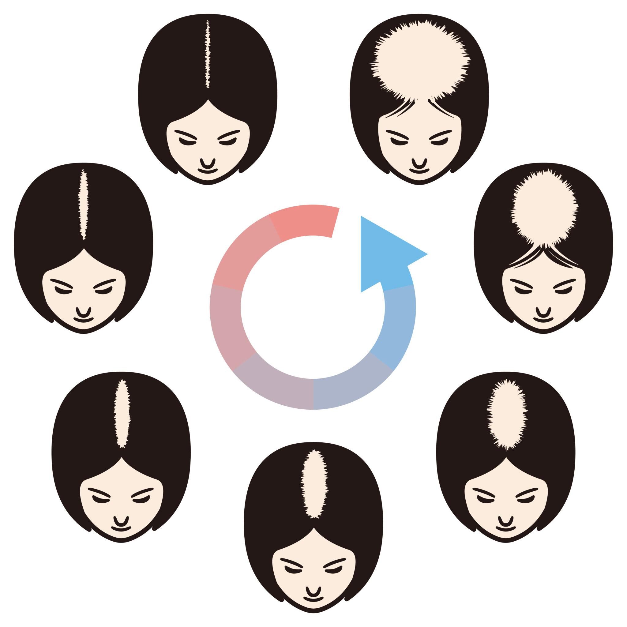
Hair moves through anagen, catagen, and telogen phases of the hair cycle, and individual hairs do not transition in synchrony.
Before-after photos taken 1–2 months after treatment onset may simply be capturing the natural fluctuation of telogen shedding and regrowth.
Efficacy assessment for stem cell conditioned media or oral medications requires at least a 3–6 month observation window, with 12-month reassessment as the standard.
When short-term case photos are presented as “dramatic change,” cross-reference them against the hair cycle timeline.
Read Whether the Initial Shedding Phase Has Been Skipped
Many hair treatments cause temporary “initial shedding” as the hair cycle resets.
Directly comparing a photo at 1–2 months with one at 6 months (after passing through initial shedding) tends to exaggerate the apparent change.
Whether progression snapshots are published continuously — including the dip in the middle — is a key indicator of whether the presentation is honest.
Cohort: Case Photos Are Biased Toward Favorable Responders
Published Cases Carry Best-Case Selection Bias
Case photos published for public use are structurally biased toward favorable responders — those who consented and whose change is visually clear.
How many of the people receiving the same treatment achieved comparable change — that is, the distribution of efficacy across the full cohort — cannot be read from a single case photo.
When comparing treatments, directly asking the physician “of how many cases is this typical” and “what is the average response” is the shortcut.
From a case photo literacy standpoint, not anchoring your expectations to the best case is essential for avoiding inflated hopes and subsequent disappointment.
Seek Cases Close to Your Own Condition
To apply the reading of change to yourself, it is realistic to reference cases close in age, sex, progression stage, and baseline follicular density.
You cannot compare Norwood II–III mild cases and VI–VII advanced cases on the same scale.
At the first visit, ask to see multiple progression photos of cases close to your own condition and confirm both the range and the limits of expected change.
Case Photo Literacy in Practice: Five Checkpoints
Five practical checkpoints to avoid taking case photos at face value:
First, whether shooting conditions (lighting, angle, distance, hair state) are uniform before and after.
Second, whether the shooting date and observation period are specifically stated.
Third, whether the treatment method, number of sessions, and concomitant medications are clearly listed.
Fourth, whether “of how many cases” and the overall responder rate are shown.
Fifth, whether cases similar to your own condition — progression, age, sex — can be selected.
Only when these five align does a case photo function as genuine “decision material.”
For related information on hair regenerative medicine, see our full column index. For AGA treatment guidelines, see the Japanese Dermatological Association.
Frequently Asked Questions
Q. Can I take case photos on official websites at face value?
Even if the photos themselves are unretouched, when there is no annotation about shooting conditions, observation period, or cohort, the information density is low. Trustworthy clinics clearly state shooting protocols and observation periods. From a case photo literacy perspective, treating unannotated case photos as “reference only” is the safer stance.
Q. How should I handle case photos I see on social media?
SNS case photos are easily affected by lighting, angle, and editing apps; even when the subject is the same person, comparison precision is limited. If using them as decision material, always cross-check against the clinic’s own documentation and verify actual progression photos during consultation.
Q. Are clinics with few case photos untrustworthy?
More important than photo count is whether shooting conditions are standardized and observation periods are specified. Even with few photos, a clinic with clear protocols and honest explanations is trustworthy. Judge by the quality of information, not the quantity of photos.
Q. When documenting my own progression, what should I pay attention to?
The basics: shoot under the same lighting, same angle, same distance, same hair wetness, and at the same time of day. Keeping fixed-point photos from multiple angles before treatment enables objective evaluation at 6 and 12 months. Even a smartphone can achieve adequate standardization with a tripod and marked position.
──────────────
Supervising Physician: Shin Moriwaki, MD
Member, Japan Society of Aesthetic Surgery (JSAS)
Member, American Academy of Aesthetic Medicine
ECFMG Certificate
──────────────
📍AVAN TOKYO Ginza Hair Regenerative Medicine
English / 中文 / Tiếng Việt available
Inquiries welcome via DM / LINE / Website / Phone.