


Measuring Hair Treatment Outcomes: Standardized Photography, Trichoscopy and Hair Diameter2026.10.16
“I look in the mirror and I can’t tell if anything has changed.” “People around me say it’s gotten thicker, but I’m not sure.” Measuring hair treatment outcomes is a far more subjective domain than most patients realize. When you see your own hair every day, small gains and losses are buried in adaptation, and the impression flips with lighting, humidity, and mood. Measuring hair treatment outcomes is work that belongs to instruments and protocols, not sensation. In this article, Dr. Moriwaki walks through how to design a system of visualization built on three objective indices: standardized photography, trichoscopy, and hair diameter measurement.
Key Points of This Article
・Measuring hair treatment outcomes is done with three objective indices — standardized photography, trichoscopy, and hair diameter measurement — not by subjective judgment.
・The evaluation timeline is baseline, 3 months, 6 months, and 12 months. These intervals are built around the hair cycle.
・Unless light source, distance, angle, and hair dryness are kept constant, real change gets lost in photographic noise.
・Because hair diameter changes before overall hair volume, short-term signals are picked up through trichoscopy and hair diameter measurement.
・Relying on photos alone or images alone misses important changes. The three indices only become meaningful when combined.
Why Subjective Evaluation Alone Falls Short When Measuring Hair Treatment Outcomes
Human vision is poor at tracking continuous change. Patients who see their own hair every day fail to perceive gradual improvement or decline, and only notice the net difference months later — often having lost track of weeks of information in the process. Lighting type (daylight vs. incandescent), mirror distance, hair wetness, and the amount of styling product all shift the apparent result. Comments from family or a hair stylist are useful, but “it looks like it’s getting thicker” is a direction, not a decision input. For deciding whether to continue, modify, or stop a treatment plan, clinic-led objective evaluation is required.
Standardized Photography — The Craft of Shooting Under the Same Conditions
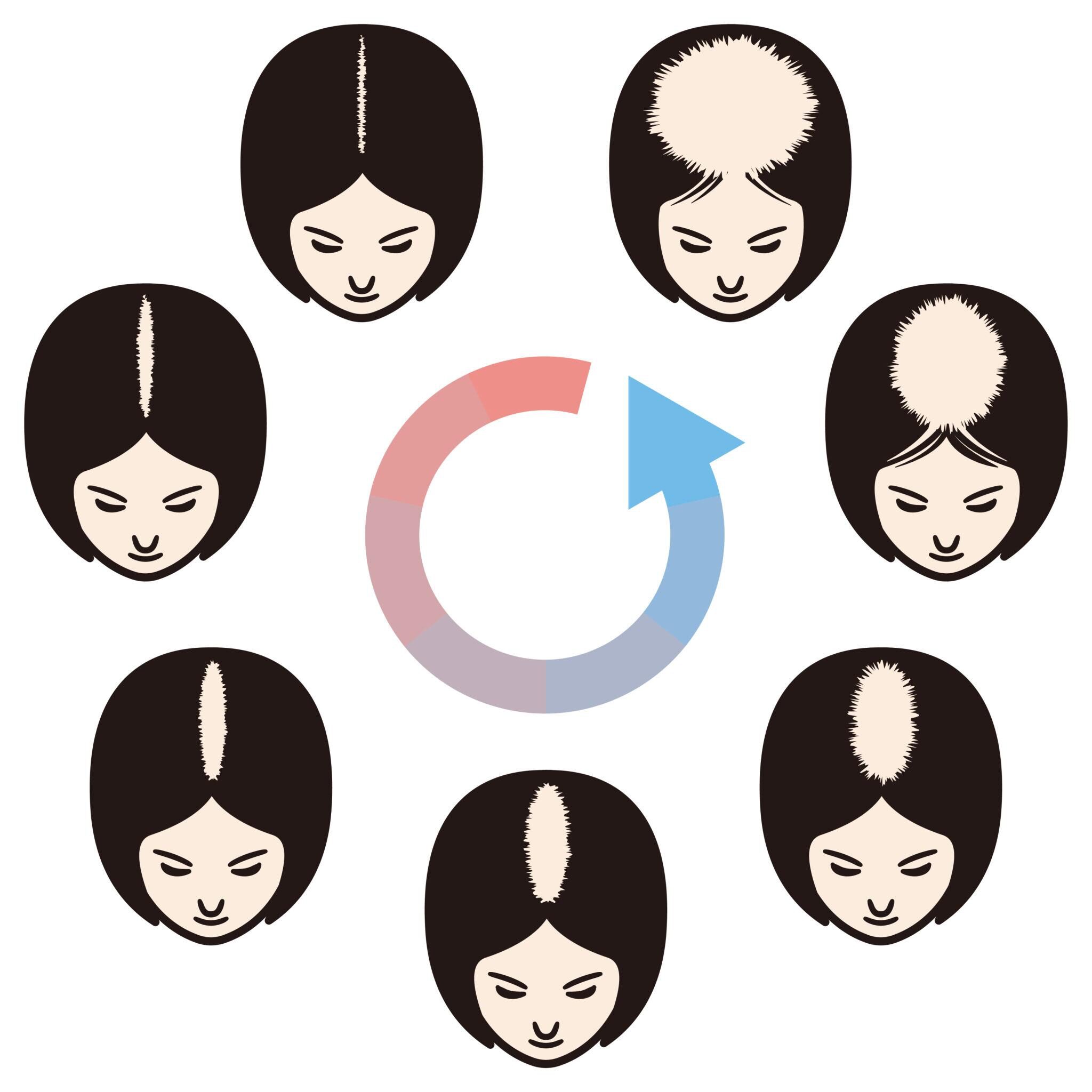
Standardized photography is the discipline of capturing images with the same conditions and the same framing at baseline and at every follow-up visit. At least five conditions need to be matched. First, light source — keep color temperature constant, and prefer stable indoor light over natural light. Second, distance — fix the distance between the camera and the subject with a tape measure. Third, angle — vertex, frontal, lateral, and occipital views, with the head oriented the same way. Fourth, hair state — same dryness, no styling product. Fifth, parting — part on the same side, or do not part the hair at all. Without these controls, a photo cannot tell you whether the change you see is in the hair or in the photo.
Trichoscopy — Visualizing Change at the Follicular Unit Level
Trichoscopy is a dermoscopic examination performed with a contact scalp dermatoscope. It inspects follicular-unit information: hairs per follicular opening, variability in hair diameter, inter-follicular spacing, peri-follicular inflammation, and the vascular pattern of the scalp. Under a successful treatment, hair diameter variability tends to normalize first, then the number of hairs per follicular opening increases, and only later does overall density rise. Trichoscopy is especially suited to answering the question that macro photography cannot: “has follicular miniaturization stopped?” Please also see our hair regeneration medicine column index.
Hair Diameter Measurement — Putting a Number on “Thickness”
Hair diameter measurement quantifies the thickness of the hair shaft in micrometers, using either harvested hairs or digital analysis of trichoscopy images. Changes in overall volume are bound by the hair cycle (the anagen phase lasts years), so three months rarely moves the volume number. Hair diameter, however, can shift over weeks to a few months. The subjective comment “I don’t feel it’s thicker yet, but it feels firmer” usually corresponds to a few microns of diameter gain, which makes hair diameter the easiest objective index to detect in the short term. For early continuation decisions, diameter data carries a disproportionate weight. For guidance on AGA treatment, you can also refer to the Japanese Dermatological Association.
Timing Design — Baseline, 3 Months, 6 Months, 12 Months
All evaluation timing is built around the hair cycle. The baseline four points are pre-treatment baseline, 3 months, 6 months, and 12 months. At 3 months we check hair diameter and the resolution of initial shedding; at 6 months we check diameter stability and change in hairs per follicular unit; at 12 months we evaluate overall density and the appearance in photos. Shorter cycles don’t give the hair cycle time to respond. Longer cycles — intervals over 12 months — delay course correction when a plan isn’t working. The right rhythm is not too fast, not too slow, matched to the hair cycle itself.
The Changes You’ll Miss If You Don’t Combine the Three Indices
Rely on standardized photos alone, and you’ll miss changes in hair diameter (firmness). Rely on trichoscopy alone, and you lose the overall-volume picture. Rely on hair diameter alone, and you can’t see uneven follicular distribution across the vertex. Captured at the same time points and cross-referenced, the three indices surface insights you can act on: “photos aren’t showing it clearly, but diameter is definitively up” or “diameter is flat, but density has climbed.” Measuring hair treatment outcomes with a single index is insufficient — this is a domain to be designed around the overlay of three.
Frequently Asked Questions
Q. Are smartphone photos taken at home useful for measuring hair treatment outcomes?
They are partially useful, but it is hard to standardize conditions, so they shouldn’t be the primary index. When position, angle, and light source vary each time, real change gets lost in the noise. If possible, continue clinic-based photography with the same device and framing.
Q. How soon after starting treatment can outcomes be evaluated?
The first checkpoint is 3 months. At this stage we look at hair diameter and the resolution of initial shedding. Visible changes in volume start to appear after 6 months, and the definitive assessment is typically made at 12 months.
Q. Is trichoscopy available at any clinic?
Specialty hair clinics or dermatology practices with the equipment generally offer it. Device models, magnifications, and image formats differ between clinics, so comparisons across clinics are harder than comparisons within the same clinic.
Q. Is it okay to decide on my own that “it’s not working” and stop?
Stopping within three months is not recommended. You may be inside an initial shedding phase, and objective indices often show the treatment is still mid-course. Please consult your doctor and review the three indices before deciding.
Q. How many micrometers of diameter increase counts as “effective”?
There is no single threshold, but a 10% or greater increase from baseline is often considered a clinically meaningful change. What matters more than the absolute number is the direction and magnitude of change over time in the same individual.
──────────────
Medical Supervisor: Shin Moriwaki, MD
Member, Japan Society of Aesthetic Surgery (JSAS) / Member, American Academy of Aesthetic Medicine
ECFMG Certificate
──────────────
📍AVAN TOKYO Ginza Hair Regenerative Medicine
AVAN TOKYO Ginza Hair Regenerative Medicine
English / 中文 / Tiếng Việt supported
Inquiries welcome via DM / LINE / Website / Phone.