


M-Pattern AGA vs. Crown-Type AGA: How Stem Cell Conditioned Media Allocation Differs at Norwood III — Dr. Moriwaki Explains2026.07.18
Even when two patients receive the same Norwood III diagnosis, is it truly reasonable to apply the same treatment plan to M-pattern AGA — where hairline recession dominates — and to crown-type AGA, where the vertex is thinning? The frontal and vertex scalp differ in follicular androgen sensitivity, blood supply, and treatment response, so reallocating oral medications, topicals, and stem cell conditioned media by pattern directly influences outcomes and patient satisfaction. In this column, Dr. Moriwaki of AVAN TOKYO Ginza (Hair Regenerative Medicine) organizes the differences between M-pattern AGA and crown-type AGA and the treatment design considerations essential at the Norwood III stage.
Key Points of This Article
・M-pattern AGA and crown-type AGA show different follicular response characteristics even at the same Norwood III stage
・The frontal scalp reacts more slowly due to 5α-reductase type II activity and limited blood flow, while the crown, being larger in area, allows earlier detection of change
・Reallocating the injection-point density and session cadence of stem cell conditioned media by pattern changes how progress becomes visible
・For combined therapy sequencing, M-pattern prioritizes hairline preservation while crown-type prioritizes area coverage
・Never decide the plan on your own — pattern classification and Norwood staging at the initial visit form the starting point of treatment design
Two Progression Patterns Within the Same Norwood III Stage
The Norwood scale describes AGA progression in seven grades, and III is the first stage where hair loss becomes clearly perceptible. Even within III, the clinical picture is not uniform: it broadly divides into M-pattern AGA (frontotemporal recession) and crown-type AGA (Norwood III vertex), with a mixed type where both coexist. Building a plan without identifying which pattern is progressing risks leaving the patient’s most-concerned area untouched, stalling satisfaction with progress.
Clinical Features of M-Pattern AGA
The temples and hairline recede, and the forehead shifts from a squared shape toward an “M” outline. Because this area strongly shapes facial impression, even a few millimeters of recession feels significant to the patient. Progression may seem slow, but recession accumulates reliably over years — many patients say, “By the time I noticed, I couldn’t turn back.”
Clinical Features of Crown-Type AGA
Hair at the vertex and crown thins and shortens, and the scalp becomes visible through the hair. Patients often fail to notice it in the mirror and become aware only through family remarks or overhead photos. Because the affected area is broad, once a threshold is crossed, patients often perceive that they “suddenly lost a lot of hair.”
Why Frontal and Vertex Scalp Respond Differently to Treatment
The rationale for separating treatment design between M-pattern AGA and crown-type AGA lies in scalp anatomy and physiology. Assuming “AGA is AGA, so the treatment is the same” causes regional differences to be missed.
5α-Reductase Distribution and Androgen Sensitivity
5α-reductase, which converts testosterone into DHT (active male hormone), shows different type I and type II distributions across the scalp. Both frontal and vertex regions are strongly influenced by type II. Follicles with high DHT sensitivity undergo shortened anagen phase and progressive miniaturization, which drives AGA progression. Finasteride and dutasteride block this pathway, but cellular response speed and remaining follicular capacity tend to be slower in the frontal scalp.
Blood Supply and Follicular Microenvironment
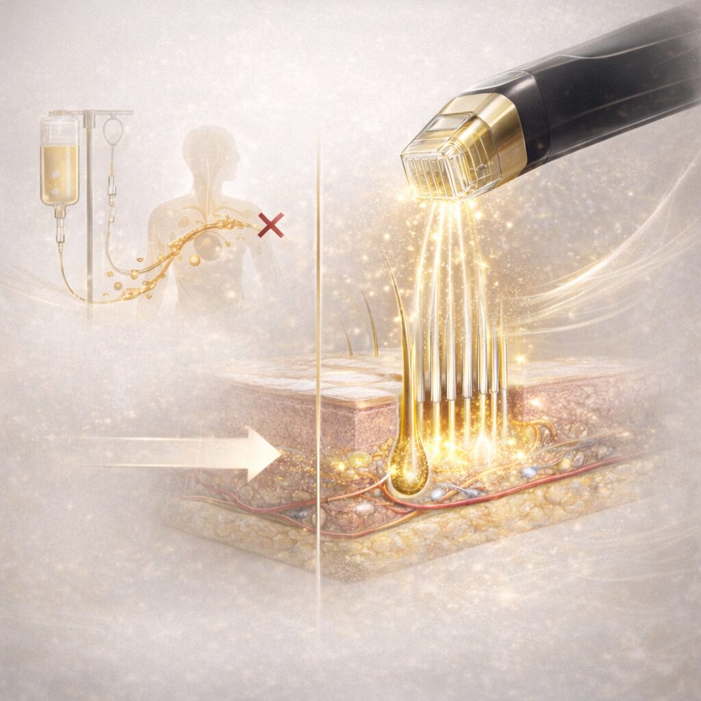
The frontal scalp has limited branches from the temporal artery, resulting in a thinner supply of oxygen and nutrients to the follicles. The vertex enjoys anastomoses from the temporal and occipital regions, giving it a relatively richer blood-flow environment. The growth-factor group delivered by stem cell conditioned media (VEGF, IGF-1, HGF, FGF, and others) is thought to act on this follicular microenvironment, transmitting signals to vessels, fibroblasts, and dermal papilla cells to help support the background against progression. However, it is not a treatment that creates lost follicles anew, so expectations must be set honestly.
How to Reallocate Stem Cell Conditioned Media for M-Pattern AGA and Crown-Type AGA
Stem cell conditioned media is not a cure-all, nor a standalone guarantee of regrowth. Clinically, it makes sense to layer it as reinforcement to the follicular microenvironment on top of a background of oral and topical therapies that slow progression. On that basis, here is how allocation shifts by pattern.
M-Pattern: Guard the Hairline as a “Line,” Not a “Surface”
Raise the injection-point density along the hairline to prioritize preventing further recession at the temples and sides. During the induction phase, deliver several sessions at 4–6 week intervals, combined with finasteride or dutasteride to suppress background progression. The frontal scalp has thin subcutaneous tissue and superficial vessels, so careful control of depth, angle, and bleeding directly shapes the finish.
Crown-Type: Prioritize Area Coverage and Envelop the Entire Vertex
Center injection points on the vertex, distributing them along the hair-flow from the temporal to the occipital region. Combining with topical minoxidil allows synergy between vasodilation and growth-factor signaling. Because area changes are visually easier to perceive, it is important to run in parallel with objective evaluation such as standardized photography, trichoscopy, and hair-diameter measurement.
Priorities for the Mixed Type (M-Pattern + Crown)
Many Norwood III cases progress in both areas simultaneously. Address the area the patient cares about most first, and avoid spreading to the full scalp all at once. Design induction and maintenance phases separately, and keep flexibility to redistribute allocation as progress is observed.
Other columns on hair regenerative medicine centered around stem cell conditioned media are available at the hair regenerative medicine column list. For AGA and alopecia clinical guidelines, please also refer to the Japanese Dermatological Association guidelines.
Frequently Asked Questions
Q. Which is harder to treat — M-pattern AGA or crown-type AGA?
Generally, the frontal region (the M-pattern area) responds more slowly in terms of blood supply and follicular reaction. However, individual variation is large, and outcomes also depend on progression stage, age, lifestyle, and when treatment began — so evaluation at the initial visit is the starting point of judgment.
Q. Should stem cell conditioned media treatment start immediately at Norwood III?
Not necessarily. In many cases, it is more rational to first establish background suppression with 5α-reductase inhibitors and minoxidil, then layer stem cell conditioned media on top as reinforcement. We will propose a treatment sequence after examination.
Q. Can conditioned media work on completely receded areas of the M-pattern?
No effect can be expected in areas where follicles have been lost and scarring has occurred. The earlier intervention takes place — while follicles still remain even at vellus-hair level — the more responsive the tissue tends to be. We check follicular remnants under a microscope before designing the plan.
Q. Is it true that crown-type AGA responds more perceptibly to treatment?
There is a tendency for density recovery to be visually easier to appreciate in a broader area. However, subjective perception and objective improvement are different, and it is important to combine evaluation axes such as photography and hair-diameter measurement.
Q. My family has AGA. Will I progress in the same pattern?
Genetic predisposition has an influence, but the progression type does not necessarily match completely. While referring to relatives’ progression, checking your own scalp to identify your pattern is the first step toward early response.
──────────────
【Medical Supervisor】Shin Moriwaki (Supervising Physician)
Member, Japan Society of Aesthetic Surgery (JSAS) / Member, American Academy of Aesthetic Medicine
ECFMG Certificate
──────────────
📍AVAN TOKYO 銀座 毛髪再生医療
AVAN TOKYO Ginza Hair Regenerative Medicine
English / 中文 / Tiếng Việt available
Inquiries welcome via DM / LINE / Website / Phone.